. 2010 Jun;38(10):3308-17.
doi: 10.1093/nar/gkq051. Epub 2010 Feb 10.

Leishmania actin binds and nicks kDNA as well as inhibits decatenation activity of type II topoisomerase

Affiliations

Leishmania actin binds and nicks kDNA as well as inhibits decatenation activity of type II topoisomerase

Prabodh Kapoor et al. Nucleic Acids Res. 2010 Jun.

Abstract

Leishmania actin (LdACT) is an unconventional form of eukaryotic actin in that it markedly differs from other actins in terms of its filament forming as well as toxin and DNase-1-binding properties. Besides being present in the cytoplasm, cortical regions, flagellum and nucleus, it is also present in the kinetoplast where it appears to associate with the kinetoplast DNA (kDNA). However, nothing is known about its role in this organelle. Here, we show that LdACT is indeed associated with the kDNA disc in Leishmania kinetoplast, and under in vitro conditions, it specifically binds DNA primarily through electrostatic interactions involving its unique DNase-1-binding region and the DNA major groove. We further reveal that this protein exhibits DNA-nicking activity which requires its polymeric state as well as ATP hydrolysis and through this activity it converts catenated kDNA minicircles into open form. In addition, we show that LdACT specifically binds bacterial type II topoisomerase and inhibits its decatenation activity. Together, these results strongly indicate that LdACT could play a critical role in kDNA remodeling.


Figures
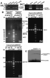
Figure 1.
Figure 1.
(A) ChIP analysis using anti-rLdACT antibodies showed the in vivo association of LdACT with chromatin and kDNA network. (a) and (b) are the agarose gels of PCR products after ChIP assay, showing the association of LdACT with nuclear DNA and kDNA, respectively. Lanes are marked on the top with their respective antibodies used in the ChIP assay and arrows indicated the genes amplified after pull down. An irrelevant, non-DNA associating antibody, GRP78, was used as a negative control, whereas, antibodies against DNA polβ, and UMSBP (universal minicircle sequence-binding protein), were used as positive controls for nuclear DNA and kDNA respectively. LdPFN, Leishmania profilin; NM12/17, specific minicircle primers. (B) Agarose gel shift assay of supercoiled and linearized pBR322 (400 ng each) in the presence of rLdACT (0.5 & 2.0 μM), and β- and γ-actins (0.5 & 2.0 μM) as indicated on the top of the gels. Lane M, shows 1 kb DNA ladder; FI: supercoiled form, FII: relaxed form, FIII: linearized form of DNA. (C) Autoradiogram of EMSA on polyacrylamide gel of 32P end-labelled 30 bp DNA probe in the presence of increasing concentration of rLdACT (0.1–0.6 μM).
Figure 2.
Figure 2.
(A) Agarose gel, showing supercoiled pBR322 DNA (400 ng) relaxation separately with rLdACT (0.5 & 1.0 μM) and E. coli Topo I. Image presented is the negative of original gel image. (B) Agarose gel electrophoresis of supercoiled pBR322 DNA (400 ng) with rLdACT in the presence or absence of anti-rLdACT antibodies, showing specificity of nicking activity associated with rLdACT. (C) Agarose gel electrophoresis of supercoiled pBR322 DNA (400 ng) with rLdACT (0.5 & 1.0 μM) in the presence or absence of DNase-1 and its inhibitor EDTA which further eliminates the possibility of DNA nicking by contaminating nuclease. (D): a and b, Agarose gel electrophoresis of supercoiled pBR322 DNA (400 ng) with rLdACT in the presence of ATP and its non-hydrolysable ATP analogs as indicated on the top of the gel. (E) Graph showing the requirement of ATP in its hydrolysable form during rLdACT mediated relaxation of supercoiled pBR322 DNA. (F) Agarose gel, showing requirement of rLdACT (0.5 & 2.0 μM) in its polymeric state for its scDNA-relaxation activity. (G) Graph, representing rLdACT (1.0 μM) mediated relaxation of supercoiled pBR322 DNA in the presence of increasing concentration of NaCl, inset shows the relative % inhibition of rLdACT mediated relaxation of supercoiled pBR322 DNA in presence of 50 mM salts having different ionization constant (Ksp) as indicated on the top of the bars. (H) Dynamic light scattering measurements of rLdACT (1.0 μM) showing insignificant effect of 0.2 M NaCl on the polymerized state of rLdACT after complete polymerization.
Figure 3.
Figure 3.
(A) Agarose gel (0.5%), showing the time dependent nicking of kDNA by rLdACT (4.0 μM) which revealed the existence of major nicked DNA and minor concatenated minicircle species. (B) Agarose gel, showing rLdACT mediated decatenation of the kDNA network in the presence or absence of anti-rLdACT antibodies. DM, decatenated kDNA marker (Topogen). (C) Agarose gel (1.0%), showing rLdACT mediated decatenation of kDNA network with rLdACT in the presence or absence of DNase-1 and its inhibitor EDTA, which completely rules out the possibility of DNA nicking by some contaminating nuclease. (D) Agarose gel (1.0%), showing requirement of rLdACT in its polymeric state for its kDNA decatenation activity. (E): (a), Agarose gel (1.0%), showing requirement of ATP in the rLdACT mediated kDNA decatenation process. (b), Graph, showing ATP dependence of rLdACT-mediated kDNA decatenation. (F) (a), Agarose gel (1.0%), showing rLdACT-mediated decatenation of kDNA in the presence of non-hydrolysable analogs of ATP. (b) Graph, showing relative inhibition of rLdACT mediated decatenation of kDNA network in the presence of non-hydrolysable ATP analogs when plotted with the increasing concentration of rLdACT.
Figure 4.
Figure 4.
AFM of kDNA carried out separately in the absence (control) or presence of rLdACT, showing the decatenation of kDNA with rLdACT. Panel a and b, control kDNA, arrows indicate catenated kDNA. Panel c and d, kDNA with rLdACT, arrowheads indicate decatenated nicked kDNA. (Scale bar: 500 nm).
Figure 5.
Figure 5.
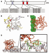
Computational docking of average simulated model of LdACT with DNA showed the interaction of the diverged DB-loop of LdACT with the major groove of DNA. (A) Sequence alignment of LdACT with other actins showed the conserved nuclear export signals (NES-1 and NES-2) and the diverged DB-loop predicted to be involved in the DNA binding, by DP-Bind server. (B) Energy minimized average simulated model of LdACT showing positions of NES-1, NES-2 (red) and the diverged stretches of amino acid sequences (yellow) including the sequence that fall in DB loop (blue). (C) Docking of LdACT (orange) with DNA (green) using HADDOCK protocols. (D) Amino acid residues of the DB loop of LdACT (yellow) showing hydrogen bonding interactions with the nucleotides (green) of DNA.
Figure 6.
Figure 6.
(A) Subtilisin mediated disruption of DB loop structure of rLdACT. a, SDS–PAGE analysis of purified rLdACT and SD-rLdACT after coomassie staining; b, western blot analysis of purified rLdACT and SD-rLdACT using anti-His6 antibodies which showed the cleavage from the N-terminus, amino acid sequences, 45–53 of rLdACT; and c, western blot analysis of purified rLdACT and SD-rLdACT using anti-rLdACT antibodies that exclude the possibility of any contamination of rLdACT in the purified SD-rLdACT fraction. (B) Model showing disruption of DB loop structure after subtilisin digestion of LdACT. (C) Agarose gel (1.0%), showing kDNA nicking with the increasing concentrations of rLdACT (0.5 – 2.0 μM) and SD-rLdACT (0.5 – 2.0 μM) as indicated on the top of the gel. (D) Bar graph, representing relative nicking of kDNA network in the presence of rLdACT and SD-rLdACT.
Figure 7.
Figure 7.
rLdACT inhibits the kDNA decatenation activity of E. coli type II topoisomerase and physically interacts with this enzyme. (A) Agarose gel (1.0%), showing nicking of kDNA in the presence or absence of rLdACT as indicated on the top of the gel. (B) SDS–PAGE showing binding of rLdACT specifically with topoisomerase-II assessed by the pulldown assay using anti-rLdACT antibodies. For these experiments, equimolar amounts of rLdACT and Topo II or Topo I were incubated in 10 mM Tris–Cl buffer (pH 7.5) containing 0.1 mM dithiothretol for 30 min. It was mixed with anti-rLdACT antibodies for 1 h and then with Protein A-Sepharose beads for 15 min. The mixture was centrifuged and the beads were washed two times and proteins absorbed on beads were eluted by SDS–PAGE sample buffer and subjected to analysis. Lanes 1–3 are the purified proteins used during the pull down experiment; Lane 1, rLdACT (43 kDa); Lane 2, Topo II (heterotetramer made up of 2 gyrA (97 kDa) subunits and 2 gyrB (90 kDa) subunits); Lane 3, Topo I (holoenzyme 97 kDa). Lanes 4–11 are the proteins after pulldown using anti-LdACT antibodies. P, proteins on protein A-sepharose beads; S, unbound proteins in the supernatant. The arrow Ab, marks the antibody band.

Similar articles

Cited by

References

    1. Hofmann WA. Cell and molecular biology of nuclear actin. Int. Rev. Cell Mol. Biol. 2009;273:219–263. - PubMed
    1. Louvet E, Percipalle P. Transcriptional control of gene expression by actin and myosin. Int. Rev. Cell Mol. Biol. 2009;272:107–147. - PubMed
    1. Farrants AK. Chromatin remodelling and actin organisation. FEBS Lett. 2008;582:2041–2050. - PubMed
    1. Liu B, Liu Y, Motyka SA, Agbo EE, Englund PT. Fellowship of the rings: the replication of kinetoplast DNA. Trends Parasitol. 2005;21:363–369. - PubMed
    1. Lukes J, Guilbride DL, Votypka J, Zikova A, Benne R, Englund PT. Kinetoplast DNA network: evolution of an improbable structure. Eukaryot. Cell. 2002;1:495–502. - PMC - PubMed

Publication types