. 2010 Feb 10;30(6):2257-67.
doi: 10.1523/JNEUROSCI.1820-09.2010.

The guanine exchange factor vav controls axon growth and guidance during Drosophila development

Affiliations

The guanine exchange factor vav controls axon growth and guidance during Drosophila development

Marianne Malartre et al. J Neurosci. .

Abstract

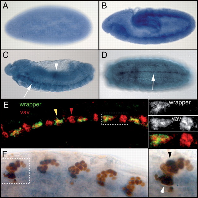
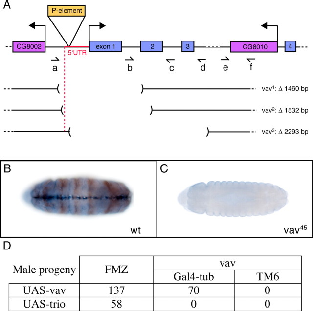
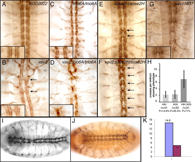
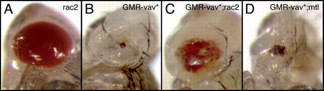
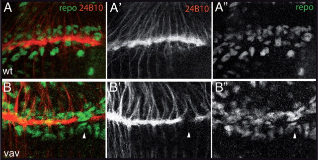
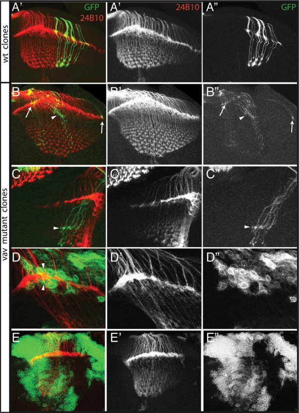
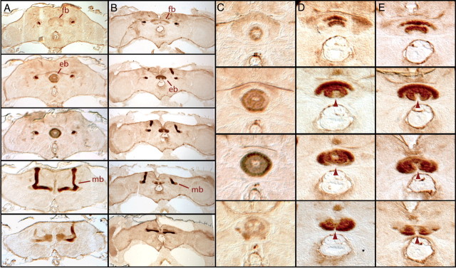
The Vav proteins are guanine exchange factors (GEFs) that trigger the activation of the Rho GTPases in general and the Rac family in particular. While the role of the mammalian vav genes has been extensively studied in the hematopoietic system and the immune response, there is little information regarding the role of vav outside of these systems. Here, we report that the single Drosophila vav homolog is ubiquitously expressed during development, although it is enriched along the embryonic ventral midline and in the larval eye discs and brain. We have analyzed the role that vav plays during development by generating Drosophila null mutant alleles. Our results indicate that vav is required during embryogenesis to prevent longitudinal axons from crossing the midline. Later on, during larval development, vav is required within the axons to regulate photoreceptor axon targeting to the optic lobe. Finally, we demonstrate that adult vav mutant escapers, which exhibit coordination problems, display axon growth defects in the ellipsoid body, a brain area associated with locomotion control. In addition, we show that vav interacts with other GEFs known to act downstream of guidance receptors. Thus, we propose that vav acts in coordination with other GEFs to regulate axon growth and guidance during development by linking guidance signals to the cytoskeleton via the modulation of Rac activity.


Figures

Figure 1.
Figure 1.
Whole-mount in situ hybridization to detect vav expression in Drosophila embryos. Anterior is toward the left in all panels. A, Vav is ubiquitously expressed in the cellular blastoderm. B, Lateral view of a stage 11 embryo when the germ band is extended, showing vav expression in the midline primordium and the head. C, Stage 13 germ band-retracted embryo. Lateral view shows vav expression in the embryonic midline (white arrow) and in the midgut (a region with higher levels of vav expression is indicated with a white arrowhead). D, Ventral view of stage 15 embryo highlighting vav expression in the midline (white arrow). E, F, Colocalization of vav mRNA and midline marker proteins in stage 15 embryos. E, Left, Confocal views of vav fluorescent in situ hybridization. Vav transcripts (red) colocalize with wrapper proteins (green), showing that vav is expressed in anterior and medial midline glia (yellow arrowhead) plus other cells where Wrapper expression is excluded (red arrowhead). Right, High magnification of region shown in the dashed rectangle. Wrapper expression alone is shown at the top, vav expression in the middle, and merged staining at the bottom. F, Left, Distribution of vav transcripts (blue) with regard to X55 enhancer-trap expression (brown) driven in midline neurons and posterior midline glia (MGP). Vav is expressed in the MGP (white arrowhead) and a subset of neurons (black arrowhead). Right, High magnification of the segment in the dashed box showing vav expression in both glial and neuronal midline cells.
Figure 2.
Figure 2.
Characterization of the vav deletion mutants. A, Schematic representation of the vav locus and mutations. Exons are represented by blue boxes, surrounding genes by purple boxes, and the P-element by an orange box. Shown in red is the vav 5′ untranslated region (UTR). Dashed lines indicate that the gene sequences continue but are not represented in entirety. Horizontal arrows designate transcription start sites for vav, for the gene located upstream and for the gene within the third exon of vav. Half-arrows (af) show the positions and directions (from 5′ to 3′) of the different primers used to determine the deletion endpoints of the three vav mutant alleles by PCR. vav1, vav2, and vav3 are the deletion mutants derived from y1P{SUPor-P}vavKG02022 by P-element imprecise excisions. The size of the deletions is indicated. For the three mutants, the vav locus is represented by a line and the region between brackets corresponds to the deletion. B, C, Vav expression is abolished in mutants. Anterior is to the left. Stage 15 embryos stained for β-gal (brown, immunostaining) and vav (blue, in situ hybridization). B, Vav2/FM7-eve-LacZ. The balancer chromosome gives positive β-gal staining and expresses the vav gene. Wt, Wild type. C, vav2 mutant shown by the absence of β-gal staining. The vav gene is not expressed. D, Table illustrating the rescue experiments. Crosses are described in Materials and Methods. The numbers of the adult males progeny are indicated in the table showing that the ubiquitous ectopic expression of vav but not trio can rescue the vav mutant lethality.
Figure 3.
Figure 3.
Vav is involved in axon formation during embryonic development and interacts genetically with other GEFs. Embryos were stained with the monoclonal anti-FasII (or 1D4) antibody to visualize a subset of CNS axons. Stage 17 embryos were filleted (B–D) or mounted in epon (A, EG). Anterior is up. Higher magnification is shown in insets. A, KG02022 embryo showing the normal organization of the longitudinal axons in three fascicles at each lateral side of the ventral midline. B, Vav2 mutant embryos showing axon formation and guidance defects. Mutants display midline crossing defects where axon bundles abnormally cross the midline (arrows). C, Trio mutants (trio6A/trio6A) display interrupted axon tracts. D, In vav;trio double mutants (vav2;trio6A/trio6A), axon tracts are more severely disrupted and axons fascicles are joined together at the midline. E, Sos embryos (sose2H/sose2H) display midline crossing defects (arrows). F, In vav;sos double mutants (vav2;sose2H/sose2H), midline crossing defects (arrows) are much stronger than in the single mutants. G, The vav 11837FRT19A mutant embryos display the same defects as the vav alleles isolated here (compare with B). H, Quantification of the midline crossing defects observed in vav, sos, and vav;sos mutants. Partial genotypes are indicated on the x-axis with the number of embryos analyzed (n) and the penetrance (P) of the phenotype in each case. The average number of ectopic crosses per embryo displaying at least one cross (expressivity) is indicated on the y-axis. Error bars represent standard deviations. I, Vav2 mutant embryos show axon defects. J, Ubiquitous expression of a wild-type version of vav, UAS-vav-HA, tagged with a HA epitope (brown), can partially rescue the embryonic CNS defects. K, Quantification of the rescue of the neuronal phenotype. The purple bar represents the percentage of vav embryos showing midline crossing defects, and the pink bar represents the rescue of this phenotype by ubiquitous expression of a wild-type version of vav.
Figure 4.
Figure 4.
Vav is required for photoreceptor axon projections. A, B, Whole-mount in situ hybridization to detect vav expression in Drosophila third instar larval eye–brain complexes. A, A′, Anterior is toward the left. In wild-type larval eye imaginal disc (A) vav is expressed ubiquitously, including in photoreceptors (PR), with higher levels observed in the morphogenetic furrow (MF) region. Vav expression is abolished in vav mutants (A′). B, Confocal view of vav fluorescent in situ hybridization. Vav expression is observed in the larval CNS, including the optic lobes (OL), with higher levels seen in the midline (arrow) and two lateral lines (arrowheads) of the ventral nerve cord. C, D, Third instar photoreceptor axon projections are visualized with anti-Chaoptin (mAb 24B10) in eye–brain complexes. Axons project from the eye disc (ed) (upper left) into the brain through the optic stalk (os). C, In wild type (wt) the R1–R6 subset of photoreceptor axons stop in the lamina neuropil (la), where their growth cones form a continuous line of staining, while R7 and R8 continue into the medulla (me), forming a topographic array. DD″, Various phenotypes can be observed in vav mutants: there are gaps in the lamina plexus (D, arrow), disruption of the typical medulla array pattern (D, asterisk), complete stalling of all axons in the lamina (D′, arrow) or increased numbers of axons entering the medulla (D″, arrowhead). E, F, R2–R5 photoreceptor axons are visualized selectively using a ro-tau-lacZ reporter to assess targeting to the lamina. E, In wild type, all R2–R5 axons stop in the lamina neuropil (arrow). F, In vav mutants, some R2–R5 axons display a lamina bypass phenotype (arrowhead).
Figure 5.
Figure 5.
Differences in the ability of Rac proteins to rescue a vav gain-of-function phenotype in the eye. Adult eyes from flies with different genotypes are shown. A, GMR/+;rac2/rac2 flies do not display eye phenotype. B, GMR/vav*;rac2/+ flies showing a severe eye phenotype. C, GMR/vav*;rac2/rac2 flies display a mild eye phenotype. D, GMR/vav*;mtl/mtl flies display an eye phenotype almost similar as that in B.
Figure 6.
Figure 6.
Lamina glia differentiation and migration occur normally in vav mutant optic lobes. Preparations of wild-type (wt) and vav mutant eye–brain complexes dissected from third instar larvae. Laser confocal microscopy 2 μm sections of optic lobes in which glial cells and R cell axons were visualized with anti-repo antibody (green) and mAb 24B10 (red), respectively. A, In wild-type larvae, glial cells, which have already migrated at this stage, are organized in three layers at the lamina plexus. R cell axons target between two layers of glial cells. B, In vav mutants, glia differentiation and migration are not compromised. In addition, the number of glial cells is not reduced and the lamina regions lacking axon innervation do not correlate with a lack of glial cells (arrowheads).
Figure 7.
Figure 7.
Vav function is required in R cell axons but not in the target to control axonal projections. A–E, Laser confocal microscopy of double-immunolabeled wild-type (wt) (A) and vav mutant (B–E) mosaic late third instar visual systems is shown. R cell axons were visualized with mAb 24B10 (red), and MARCM clones are visualized with GFP. Stacks of various 1 μm sections were made. A, In wild type, GFP-marked R cell axons target to appropriate locations in the lamina and medulla. B, C, While some homozygous vav mutant R cell axons target the lamina correctly (arrows), other axons (presumably R1–R6) project to a wrong layer below the lamina (arrowhead), from where some of them (presumably R7 and R8) extend deeper in the medulla. D, Wild-type R cell axons project correctly in the lamina plexus between two layers of vav mutant epithelial and marginal glial cells. E, Large clones of vav mutant cells in the medulla and the lamina do not affect R cell axon projections.
Figure 8.
Figure 8.
Central brain defects in the adult vav mutant. Serial 7 μm paraffin frontal sections of wild-type and vav mutant adult brains were stained with the axon marker FasII to highlight different neuropilar structures. Dorsal is above. A, In wild-type larvae, central complex regions such as the fan-shaped body (fb) and the ellipsoid body (eb) are stained with FasII, as well as the mushroom bodies (mb). B, The main brain structures are present in vav mutant brains. C, Higher magnification of wild-type ellipsoid body showing the characteristic ring-like shape of this structure. D, E, Ellipsoid bodies from vav mutants show axonal growth arrest with various expressivities (arrowheads), resulting in ventrally opened structures along the midline. In the milder cases (D) only the outside ring of the ellipsoid body is opened, whereas in stronger cases (E) axons forming both the outside and inside rings are affected.

Similar articles

Cited by

References

    1. Aoki K, Nakamura T, Fujikawa K, Matsuda M. Local phosphatidylinositol 3,4,5-trisphosphate accumulation recruits Vav2 and Vav3 to activate Rac1/Cdc42 and initiate neurite outgrowth in nerve growth factor-stimulated PC12 cells. Mol Biol Cell. 2005;16:2207–2217. - PMC - PubMed
    1. Awasaki T, Saito M, Sone M, Suzuki E, Sakai R, Ito K, Hama C. The Drosophila trio plays an essential role in patterning of axons by regulating their directional extension. Neuron. 2000;26:119–131. - PubMed
    1. Bateman J, Shu H, Van Vactor D. The guanine nucleotide exchange factor trio mediates axonal development in the Drosophila embryo. Neuron. 2000;26:93–106. - PubMed
    1. Betz R, Sandhoff K, Fischer KD, van Echten-Deckert G. Detection and identification of Vav1 protein in primary cultured murine cerebellar neurons and in neuroblastoma cells (SH-SY5Y and Neuro-2a) Neurosci Lett. 2003;339:37–40. - PubMed
    1. Boquet I, Hitier R, Dumas M, Chaminade M, Préat T. Central brain postembryonic development in Drosophila: implication of genes expressed at the interhemispheric junction. J Neurobiol. 2000a;42:33–48. - PubMed

Publication types

MeSH terms

Substances