

A genetic model of chronic rhinosinusitis-associated olfactory inflammation reveals reversible functional impairment and dramatic neuroepithelial reorganization
- PMID: 20147558
- PMCID: PMC2957830
- DOI: 10.1523/JNEUROSCI.4507-09.2010
A genetic model of chronic rhinosinusitis-associated olfactory inflammation reveals reversible functional impairment and dramatic neuroepithelial reorganization
Abstract
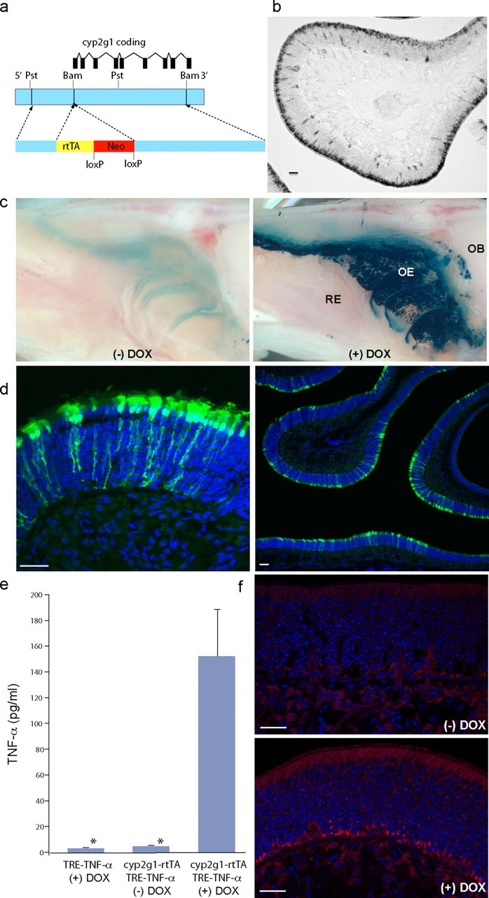
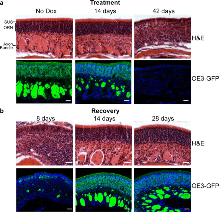
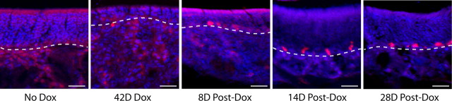
Inflammatory sinus and nasal disease is a common cause of human olfactory loss. To explore the mechanisms underlying rhinosinusitis-associated olfactory loss, we have generated a transgenic mouse model of olfactory inflammation, in which tumor necrosis factor alpha (TNF-alpha) expression is induced in a temporally controlled manner specifically within the olfactory epithelium (OE). Like the human disease, TNF-alpha expression leads to a progressive infiltration of inflammatory cells into the OE. Using this model, we have defined specific phases of the pathologic process. An initial loss of sensation without significant disruption is observed, followed by a striking reorganization of the sensory neuroepithelium. An inflamed and disrupted state is sustained chronically by continued induction of cytokine expression. After prolonged maintenance in a deficient state, there is a dramatic recovery of function and a normal histologic appearance when TNF-alpha expression is extinguished. Although obstruction of airflow is also a contributing factor in human rhinosinusitis, this in vivo model demonstrates for the first time that direct effects of inflammation on OE structure and function are important mechanisms of olfactory dysfunction. These features mimic essential aspects of chronic rhinosinusitis-associated olfactory loss, and illuminate underlying cellular and molecular aspects of the disease. This manipulable model also serves as a platform for developing novel therapeutic interventions.
Figures

Similar articles
-
The role of TNF-α in inflammatory olfactory loss.Laryngoscope. 2011 Nov;121(11):2481-6. doi: 10.1002/lary.22190. Epub 2011 Aug 31. Laryngoscope. 2011. PMID: 21882204 Free PMC article.
-
Regulation of inflammation-associated olfactory neuronal death and regeneration by the type II tumor necrosis factor receptor.Int Forum Allergy Rhinol. 2013 Sep;3(9):740-7. doi: 10.1002/alr.21187. Epub 2013 Jun 3. Int Forum Allergy Rhinol. 2013. PMID: 23733314 Free PMC article.
-
Tumor necrosis factor alpha inhibits olfactory regeneration in a transgenic model of chronic rhinosinusitis-associated olfactory loss.Am J Rhinol Allergy. 2010 Sep-Oct;24(5):336-40. doi: 10.2500/ajra.2010.24.3498. Am J Rhinol Allergy. 2010. PMID: 21243089 Free PMC article.
-
Structures and functions of the normal and injured human olfactory epithelium.Front Neural Circuits. 2024 Jun 6;18:1406218. doi: 10.3389/fncir.2024.1406218. eCollection 2024. Front Neural Circuits. 2024. PMID: 38903957 Free PMC article. Review.
-
Chronic sinusitis and olfactory dysfunction.Otolaryngol Clin North Am. 2004 Dec;37(6):1143-57, v-vi. doi: 10.1016/j.otc.2004.06.003. Otolaryngol Clin North Am. 2004. PMID: 15563907 Review.
Cited by
-
Olfactory dysfunction: its early temporal relationship and neural correlates in the pathogenesis of Alzheimer's disease.J Neural Transm (Vienna). 2015 Oct;122(10):1475-97. doi: 10.1007/s00702-015-1404-6. Epub 2015 May 6. J Neural Transm (Vienna). 2015. PMID: 25944089 Review.
-
Olfactory cleft evaluation: a predictor for olfactory function in smell-impaired patients?Eur Arch Otorhinolaryngol. 2018 May;275(5):1129-1137. doi: 10.1007/s00405-018-4913-8. Epub 2018 Feb 27. Eur Arch Otorhinolaryngol. 2018. PMID: 29488006 Clinical Trial.
-
Post Viral Olfactory Dysfunction After SARS-CoV-2 Infection: Anticipated Post-pandemic Clinical Challenge.Indian J Otolaryngol Head Neck Surg. 2022 Dec;74(Suppl 3):4571-4578. doi: 10.1007/s12070-021-02730-6. Epub 2021 Jul 7. Indian J Otolaryngol Head Neck Surg. 2022. PMID: 34249668 Free PMC article.
-
Superior turbinate eosinophilia correlates with olfactory deficit in chronic rhinosinusitis patients.Laryngoscope. 2017 Oct;127(10):2210-2218. doi: 10.1002/lary.26555. Epub 2017 Mar 21. Laryngoscope. 2017. PMID: 28322448 Free PMC article.
-
Chlamydia muridarum Can Invade the Central Nervous System via the Olfactory and Trigeminal Nerves and Infect Peripheral Nerve Glial Cells.Front Cell Infect Microbiol. 2021 Jan 8;10:607779. doi: 10.3389/fcimb.2020.607779. eCollection 2020. Front Cell Infect Microbiol. 2021. PMID: 33489937 Free PMC article.
References
-
- Aiba T, Nakai Y. Influence of experimental rhino-sinusitis on olfactory epithelium. Acta Otolaryngol. 1991;486(Suppl):184–192. - PubMed
-
- Arnett HA, Mason J, Marino M, Suzuki K, Matsushima GK, Ting JP. TNF alpha promotes proliferation of oligodendrocyte progenitors and remyelination. Nat Neurosci. 2001;4:1116–1122. - PubMed
-
- Barker V, Middleton G, Davey F, Davies AM. TNFalpha contributes to the death of NGF-dependent neurons during development. Nat Neurosci. 2001;4:1194–1198. - PubMed
-
- Caggiano M, Kauer JS, Hunter DD. Globose basal cells are neuronal progenitors in the olfactory epithelium: a lineage analysis using a replication-incompetent retrovirus. Neuron. 1994;13:339–352. - PubMed
-
- Costanzo RM, Graziadei PP. A quantitative analysis of changes in the olfactory epithelium following bulbectomy in hamster. J Comp Neurol. 1983;215:370–381. - PubMed
Publication types
MeSH terms
Substances
Grants and funding
LinkOut - more resources
Full Text Sources
Other Literature Sources
Medical
Molecular Biology Databases