. 2010 Feb 10;30(6):2330-9.
doi: 10.1523/JNEUROSCI.5574-09.2010.

Mechanisms underlying lateral GABAergic feedback onto rod bipolar cells in rat retina

Affiliations

Mechanisms underlying lateral GABAergic feedback onto rod bipolar cells in rat retina

Andrés E Chávez et al. J Neurosci. .

Abstract

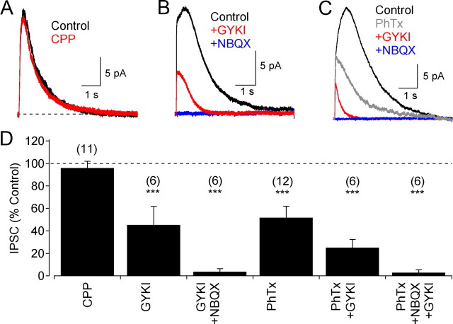
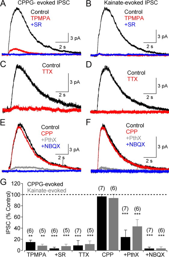
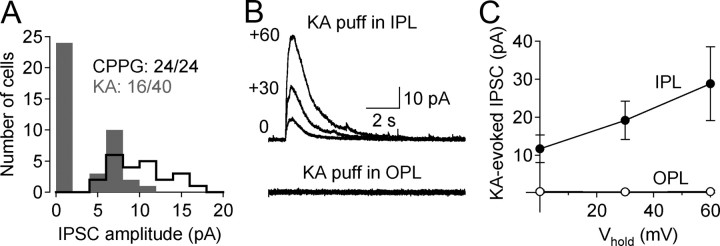
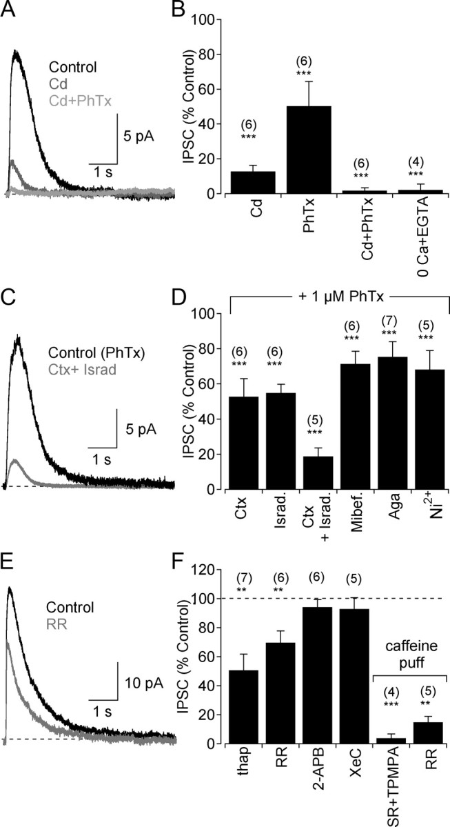
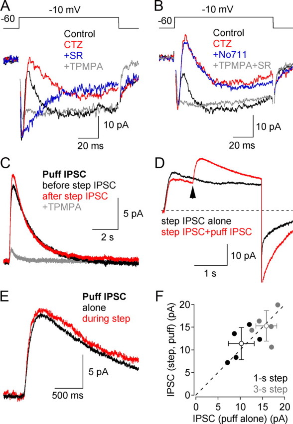
GABAergic feedback inhibition from amacrine cells shapes visual signaling in the inner retina. Rod bipolar cells (RBCs), ON-sensitive cells that depolarize in response to light increments, receive reciprocal GABAergic feedback from A17 amacrine cells and additional GABAergic inputs from other amacrine cells located laterally in the inner plexiform layer. The circuitry and synaptic mechanisms underlying lateral GABAergic inhibition of RBCs are poorly understood. A-type and rho-subunit-containing (C-type) GABA receptors (GABA(A)Rs and GABA(C)Rs) mediate both forms of inhibition, but their relative activation during synaptic transmission is unclear, and potential interactions between adjacent reciprocal and lateral synapses have not been explored. Here, we recorded from RBCs in acute slices of rat retina and isolated lateral GABAergic inhibition by pharmacologically ablating A17 amacrine cells. We found that amacrine cells providing lateral GABAergic inhibition to RBCs receive excitatory synaptic input mostly from ON bipolar cells via activation of both Ca(2+)-impermeable and Ca(2+)-permeable AMPA receptors (CP-AMPARs) but not NMDA receptors (NMDARs). Voltage-gated Ca(2+) (Ca(v)) channels mediate the majority of Ca(2+) influx that triggers GABA release, although CP-AMPARs contribute a small component. The intracellular Ca(2+) signal contributing to transmitter release is amplified by Ca(2+)-induced Ca(2+) release from intracellular stores via activation of ryanodine receptors. Furthermore, lateral nonreciprocal feedback is mediated primarily by GABA(C)Rs that are activated independently from receptors mediating reciprocal feedback inhibition. These results illustrate numerous physiological differences that distinguish GABA release at reciprocal and lateral synapses, indicating complex, pathway-specific modulation of RBC signaling.


Figures

Figure 1.
Figure 1.
Spatial profile of reciprocal and lateral feedback inhibition to RBCs. A, GABAergic IPSCs evoked by glutamate puffed at incremental distances from patched RBC (Vhold = 0 mV) were locally sensitive to DHT (50 μm) with the remaining component blocked by TTX (0.5 μm). B, Same experiment as in A, but with reversed pharmacological application (TTX first). C, D, Summary of pharmacological block at of GABAergic IPSCs as a function of lateral distance from the inhibited RBCs (n = 6). E, Addition of the average TTX-sensitive and DHT-sensitive current amplitudes closely matched the average control responses indicating that signaling from two, independent sources mediated the total response. F, Inset, In the presence of DHT, feedback IPSCs were strongly reduced by application of TPMPA (50 μm), a GABACR antagonist, and eliminated by additional inclusion of SR95531 (10 μm), a GABAAR antagonist. F, Summarized drug effects (mean ± SD) on puff-evoked feedback IPSCs. All experiments were conducted in the presence of strychnine (3 μm) to block lateral inhibition from GlyRs (Chávez and Diamond, 2008). ***p < 0.001. SR, SR95531; CGP, CGP54266.
Figure 2.
Figure 2.
Non-NMDARs mediate excitatory inputs to lateral GABAergic amacrine cells. A, Lateral GABAergic feedback IPSCs were insensitive to the NMDAR antagonist, CPP (10 μm). B, The responses were partially reduced by application of the AMPAR antagonist, GYKI (50 μm), and eliminated by coapplication with NBQX (25 μm). C, The CP-AMPAR antagonist, PhTx (1 μm), partially reduced feedback IPSCs as did GYKI; the remaining response was eliminated by additional application of NBQX (25 μm). D, Summarized drug effects (mean ± SD) on lateral GABAergic feedback IPSCs. All experiments were conducted in the presence of strychnine (3 μm) and DHT (50 μm). ***p < 0.001.
Figure 3.
Figure 3.
ON and OFF retinal pathways trigger lateral inhibition from GABAergic amacrine cells. A, Feedback IPSCs elicited by activation of ON bipolar cell dendrites (puff application of the mGluR antagonist CPPG; 600 μm) in the OPL were strongly reduced by TPMPA (50 μm) and reduced further by SR95531 (10 μm). B, Similar results were observed when OFF bipolar cell dendrites were activated by brief puffs of kainate (100 μm). C, D, “ON” (CPPG-evoked) (C) and “OFF” (kainate-evoked) (D) responses were eliminated by TTX (0.5 μm). E, CPPG-evoked IPSCs were unaffected by CPP (10 μm) but were strongly reduced by PhTx (1 μm) and eliminated by NBQX (25 μm). F, Kainate-evoked IPSCs also were insensitive to CPP (10 μm), strongly reduced by PhTx (1 μm), and eliminated by NBQX (25 μm). G, Summarized drug effects (mean ± SD) on feedback IPSCs evoked by CPPG (black bars) and kainate (gray bars). All experiments were conducted in the presence of strychnine (3 μm), DHT (50 μm), and l-AP-4 (10 μm). **p < 0.01; ***p < 0.001.
Figure 4.
Figure 4.
Lateral GABAergic feedback onto RBCs is driven more strongly through the ON pathway. A, Amplitude histogram comparing CPPG- and kainate-evoked feedback IPSCs (OPL puff application). B, Sometimes kainate failed to evoke IPSCs when puffed directly into the OPL (bottom panel) despite robust responses that could be produced by placing the same puff pipette in the IPL to activate amacrine cell dendrites directly (n = 18). C, Summarized data (mean ± SD) from experiments illustrated in B. All experiments were performed in the presence of strychnine (3 μm), DHT (50 μm), and l-AP-4 (10 μm).
Figure 5.
Figure 5.
Calcium signals underlying GABA release during lateral feedback. A, Glutamate-evoked lateral feedback IPSCs were strongly reduced by the nonselective Cav channel blocker, Cd2+ (200 μm). The Cd2+-insensitive component of the IPSCs was eliminated by additional inclusion of PhTx (1 μm). B, Summarized drug effects (mean ± SD) on feedback IPSCs. C, Feedback IPSCs evoked in the presence of PhTx (to eliminate Ca2+ influx through AMPARs; control trace) were strongly, but not completely, reduced by coapplication of either N- or L-type Cav channel antagonists (ω-conotoxin GVIA, 10 nm, or isradipine, 10 μm, respectively). D, Summarized effects of Cav channel blockers (mean ± SD) on feedback IPSCs evoked in the presence of 1 μm PhTx. E, Feedback IPSCs were reduced by bath application of the RyR antagonist, RR (40 μm), but not the IP3R antagonist XeC (3 μm). F, Summarized data (mean ± SD) showing that RyRs, but not IP3Rs, contribute to the Ca2+ signaling underlying lateral GABA release. All experiments were performed in the presence of strychnine (3 μm) and DHT (50 μm). **p < 0.01; ***p < 0.001. Israd, Isradipine; Mibef, mibefradil; Aga, agatoxin IVA; thap, thapsigargin.
Figure 6.
Figure 6.
GABACR populations activated at lateral and reciprocal feedback synapses are distinct. A, Depolarizing voltage steps (50 mV) elicited reciprocal feedback IPSCs (vIPSCs) (black trace) that were enhanced by blocking AMPAR desensitization with CTZ (50 μm; red trace). Additional application of SR95531 (10 μm; blue trace) blocked a transient component of the response and revealed a slow GABACR-mediated component that was eliminated by TPMPA (50 μm; gray trace). B, NO-711 (10 μm; blue trace) did not further enhance the GABACR-mediated component of the vIPSC. Experiments shown in A and B were done in the presence of TTX and absence of DHT. C, Example from GABACR occlusion experiments: GABACR-mediated nonreciprocal puff-evoked feedback IPSCs (stimulated ∼60–80 μm from the inhibited RBC) were elicited alone (black) or directly after (red) step-evoked reciprocal activation of GABACRs. These experiments were conducted in the presence of CTZ (50 μm) and SR95531 (10 μm) and in the absence of TTX and DHT. D, vIPSCs showed a slow and sustained GABACR-mediated component IPSC (black trace) that was not occluded by concurrent puff activation of lateral GABAergic feedback synapses (red trace; arrow indicates puff onset). E, Comparison of control puff-evoked lateral inhibition from C and subtraction of traces in D suggest that distinct GABACR populations are involved in reciprocal versus lateral GABAergic feedback. F, Summarized data (mean ± SD) from GABACR occlusion experiments presented in C–E. These experiments (C–E) were conducted in the absence of TTX and DHT.

Similar articles

Cited by

References

    1. Bieda MC, Copenhagen DR. N-type and L-type calcium channels mediate glycinergic synaptic inputs to retinal ganglion cells of tiger salamanders. Vis Neurosci. 2004;21:545–550. - PMC - PubMed
    1. Bloomfield SA. Relationship between receptive and dendritic field size of amacrine cells in the rabbit retina. J Neurophysiol. 1992;68:711–725. - PubMed
    1. Bloomfield SA, Völgyi B. Response properties of a unique subtype of wide-field amacrine cell in the rabbit retina. Vis Neurosci. 2007;24:459–469. - PubMed
    1. Bloomfield SA, Xin D. Surround inhibition of mammalian AII amacrine cells is generated in the proximal retina. J Physiol. 2000;523:771–783. - PMC - PubMed
    1. Boos R, Schneider H, Wässle H. Voltage- and transmitter-gated currents of all-amacrine cells in a slice preparation of the rat retina. J Neurosci. 1993;13:2874–2888. - PMC - PubMed

Publication types