. 2010 Feb 10;30(6):2396-405.
doi: 10.1523/JNEUROSCI.3843-09.2010.

A subpopulation of neuronal M4 muscarinic acetylcholine receptors plays a critical role in modulating dopamine-dependent behaviors

Affiliations

A subpopulation of neuronal M4 muscarinic acetylcholine receptors plays a critical role in modulating dopamine-dependent behaviors

Jongrye Jeon et al. J Neurosci. .

Abstract

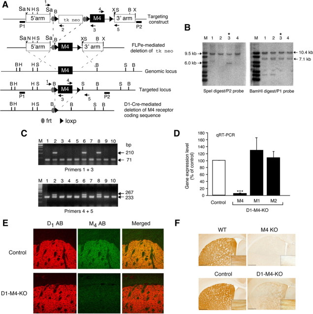
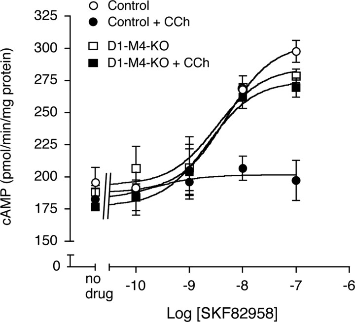
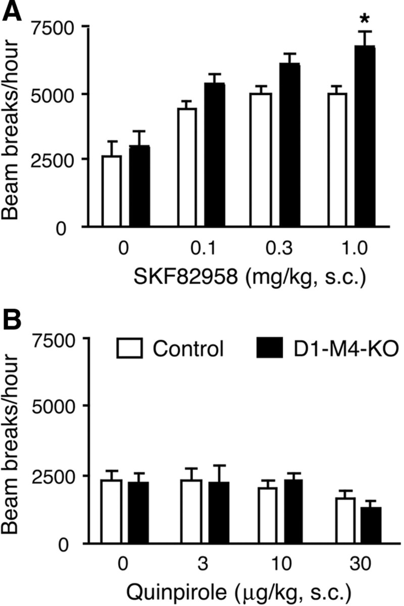
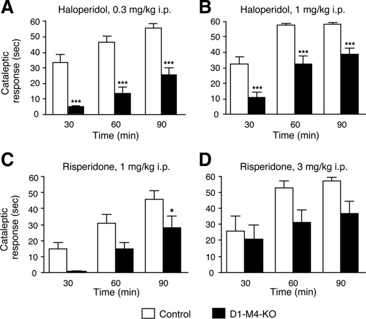
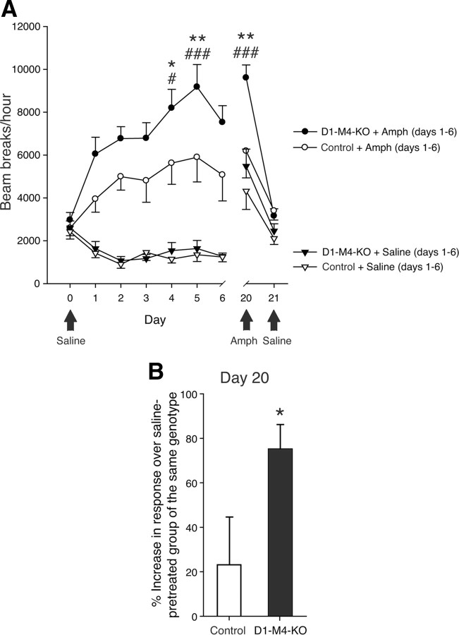
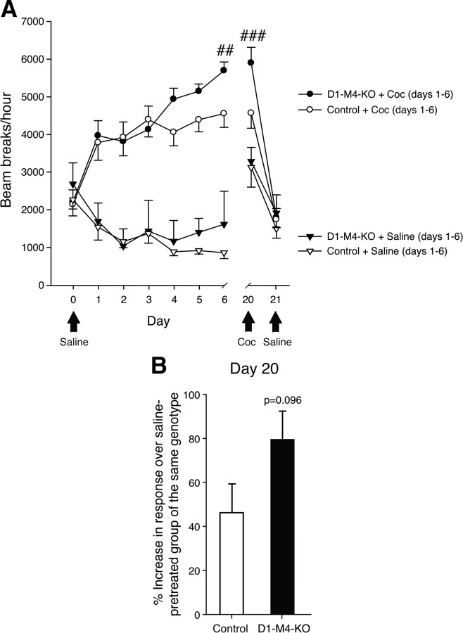
Acetylcholine (ACh) regulates many key functions of the CNS by activating cell surface receptors referred to as muscarinic ACh receptors (M(1)-M(5) mAChRs). Like other mAChR subtypes, the M(4) mAChR is widely expressed in different regions of the forebrain. Interestingly, M(4) mAChRs are coexpressed with D(1) dopamine receptors in a specific subset of striatal projection neurons. To investigate the physiological relevance of this M(4) mAChR subpopulation in modulating dopamine-dependent behaviors, we used Cre/loxP technology to generate mutant mice that lack M(4) mAChRs only in D(1) dopamine receptor-expressing cells. The newly generated mutant mice displayed several striking behavioral phenotypes, including enhanced hyperlocomotor activity and increased behavioral sensitization following treatment with psychostimulants. These behavioral changes were accompanied by a lack of muscarinic inhibition of D(1) dopamine receptor-mediated cAMP stimulation in the striatum and an increase in dopamine efflux in the nucleus accumbens. These novel findings demonstrate that a distinct subpopulation of neuronal M(4) mAChRs plays a critical role in modulating several important dopamine-dependent behaviors. Since enhanced central dopaminergic neurotransmission is a hallmark of several severe disorders of the CNS, including schizophrenia and drug addiction, our findings have substantial clinical relevance.


Figures

Figure 1.
Figure 1.
Strategy used for the conditional deletion of the M4 mAChR gene in D1 dopamine receptor-expressing neurons. A, Partial restriction maps of the wild-type (WT) M4 mAChR genomic locus, targeting construct, recombinant targeted allele, and M4 mAChR-deleted locus. The approximate locations of the frt and loxP sites, the P1 and P2 probes used for Southern analysis (filled bars), and the primers (arrows) used for PCR genotyping studies are indicated. H, HindIII; N, NotI; B, BamHI; X, XhoI; S, SpeI; Sa, SacII. B, Southern blot analysis of genomic DNA from representative ES cell clones before FLPe-mediated deletion of the tk/neo cassette. SpeI- or BamHI-digested DNA was analyzed with the P2 and P1 probes, respectively. The 9.5 and 10.4 kb bands indicate the presence of the WT M4 receptor allele, whereas the 6.0 and 7.1 kb bands are diagnostic for the proper integration of the targeting construct. Note that ES cell clone 3 (marked with an asterisk) showed the proper targeting event. C, PCR genotyping analysis of ES cell DNA after FLPe-mediated deletion of the tk/neo cassette. The 71 and 210 kb bands indicate the presence of the WT and the floxed M4 receptor allele, respectively (use of primers 1 and 3, top). Analogously, the 233 and 267 bp bands are diagnostic for the presence of the WT and the floxed M4 receptor allele, respectively (use of primers 4 and 5, bottom). Note that ES cell clones 2, 6, and 8 were heterozygous for the floxed M4 receptor allele. The same strategy was used for the genotyping of mouse tail DNA. D, Real-time qRT-PCR analysis of M1, M2, and M4 mAChR mRNA expression in the striatum of control mice (M4 fl/fl) and D1–M4-KO (M4 fl/fl D1-Cre) mice. Data were normalized by the expression of cyclophilin A, which served as an internal control. Results are shown as relative gene expression levels compared to control mice (100%; means ± SEM; n = 4 per group). ***p ≤ 0.001 versus control. E, Confocal microscopy analysis of M4 mAChR and D1 dopamine receptor expression in the striatum. The three top panels demonstrate that M4 muscarinic and D1 dopamine receptor proteins were colocalized in the striatum of control mice. The three bottom panels indicate that striatal M4 mAChR staining was reduced to background levels in D1–M4-KO mice. F, Immunohistochemical localization of M4 mAChRs in selected brain regions. As expected, M4 mAChR staining was abolished throughout the brain in whole-body M4 receptor KO mice, indicative of the specificity of the M4 mAChR antibody used (top right). The corresponding WT mice gave the expected M4 mAChR staining pattern (top left). In brain sections from WT (control) mice (left), M4 mAChR staining was abundant in the striatum and in presumed axonal processes in the corpus callosum (high-power inset). In the corresponding sections from D1–M4-KO mice (bottom right), M4 mAChR staining was greatly reduced in the striatum, but was retained in other areas of the brain such as the corpus callosum (inset).
Figure 2.
Figure 2.
Muscarinic inhibition of D1 dopamine receptor-stimulated cAMP accumulation is abolished in the striatum of D1–M4-KO mice. cAMP assays were performed using striatal membranes prepared from D1–M4-KO and control mice by applying increasing concentrations of the D1 receptor agonist, SKF82958, either in the absence or in the presence of 100 μm carbachol (CCh). Data are given as means ± SEM (n = 3).
Figure 3.
Figure 3.
Effects of SKF82958 and quinpirole on locomotor activity in D1–M4-KO and control mice. A, B, D1–M4-KO mice and control littermates were injected subcutaneously with the indicated doses of the D1 receptor agonist, SKF82958 (A), or the D2 receptor agonist, quinpirole (B). Locomotor activity was assessed during a 1 h test period. Data are given as means ± SEM (n = 9–12). *p ≤ 0.05 versus control.
Figure 4.
Figure 4.
Effects of amphetamine and cocaine on locomotor activity in D1–M4-KO and control mice. A, B, D1–M4-KO mice and control littermates were injected subcutaneously with the indicated doses of amphetamine (A) or cocaine (B). Locomotor activity was assessed during a 1 h test period. Data are given as means ± SEM (n = 9–12). **p < 0.01 versus control.
Figure 5.
Figure 5.
Cataleptic responses to haloperidol and risperidone in D1–M4-KO and control mice. A–D, D1–M4-KO mice and control littermates were injected intraperitoneally with the indicated doses of haloperidol (A, B) or risperidone (C, D), and cataleptic responses were assessed 30, 60 and 90 min after drug injection. Significant genotype effects were found at both doses of haloperidol and risperidone, indicating that D1–M4-KO mice showed attenuated cataleptic responses as compared to control mice. Data are given as means ± SEM (n = 8–18). *p < 0.05, ***p < 0.001 versus control.
Figure 6.
Figure 6.
Behavioral sensitization after repeated amphetamine injections in D1–M4-KO mice. A, Locomotor activity during sensitization to amphetamine (2 mg/kg, i.p.). Repeated administration of amphetamine significantly increased the hyperlocomotor response of D1–M4-KO mice. Black arrows indicate treatments that differed from those indicated by the legend. *p < 0.05, **p < 0.01 amphetamine-treated D1–M4-KO versus amphetamine-treated control mice; #p < 0.05, ###p < 0.001 versus amphetamine response on day 1 (same genotype). B, Amphetamine-induced locomotor responses on day 20 (test day). On day 20, all mice, independent of whether they had been treated with saline or amphetamine during the initial 6 d injection period, received a single dose of amphetamine (2 mg/kg, i.p.). For each genotype, data are expressed as percentage increase in amphetamine-induced hyperlocomotor activity observed with amphetamine-pretreated versus saline-pretreated mice. This analysis revealed a significant difference between D1–M4-KO and control mice. *p < 0.05 versus control. Data are given as means ± SEM (n = 11–16).
Figure 7.
Figure 7.
Behavioral sensitization after repeated cocaine injections in D1–M4-KO mice. A, Locomotor activity during sensitization to cocaine (10 mg/kg, i.p.). Repeated administration of cocaine significantly increased the hyperlocomotor response of D1–M4-KO and control mice. Black arrows indicate when treatment differed from that implied by the legend. Overall analysis revealed a significant genotype effect. ##p < 0.01, ###p < 0.001 versus cocaine response on day 1 (same genotype). B, Cocaine-induced locomotor responses on day 20 (test day). On day 20, all mice, independent of whether they had been treated with saline or cocaine during the initial 6 d injection period, received a single dose of cocaine (10 mg/kg, i.p.). For each genotype, data are expressed as percentage increase in cocaine-induced hyperlocomotor activity observed with cocaine-pretreated versus saline-pretreated mice. This analysis revealed a trend toward increased locomotor activity in D1–M4-KO mice (versus control mice; p = 0.096). Data are given as means ± SEM (n = 11–16).
Figure 8.
Figure 8.
Basal and amphetamine-stimulated dopamine efflux in the nucleus accumbens (nAcc) of D1–M4-KO and control mice. A, Basal dopamine efflux. Basal dopamine levels were determined via in vivo microdialysis. Data are expressed as means ± SEM (n = 6 or 7) of microdialysis samples 1–3 (see Materials and Methods for details). B, Amphetamine-stimulated dopamine efflux. D1–M4-KO mice and control littermates were injected with amphetamine (2 mg/kg, s.c.) at the 90 min time point (arrow). Data are expressed as means ± SEM (n = 6 or 7). *p < 0.05 versus control; #p < 0.05 versus basal dopamine efflux.

Similar articles

Cited by

References

    1. Bernard V, Normand E, Bloch B. Phenotypical characterization of the rat striatal neurons expressing muscarinic receptor genes. J Neurosci. 1992;12:3591–3600. - PMC - PubMed
    1. Borgkvist A, Fisone G. Psychoactive drugs and regulation of the cAMP/PKA/DARPP-32 cascade in striatal medium spiny neurons. Neurosci Biobehav Rev. 2007;31:79–88. - PubMed
    1. Brady AE, Jones CK, Bridges TM, Kennedy JP, Thompson AD, Heiman JU, Breininger ML, Gentry PR, Yin H, Jadhav SB, Shirey JK, Conn PJ, Lindsley CW. Centrally active allosteric potentiators of the M4 muscarinic acetylcholine receptor reverse amphetamine-induced hyperlocomotor activity in rats. J Pharmacol Exp Ther. 2008;327:941–953. - PMC - PubMed
    1. Bymaster FP, Felder CC, Ahmed S, McKinzie D. Muscarinic receptors as a target for drugs treating schizophrenia. Curr Drug Targets CNS Neurol Disord. 2002;1:163–181. - PubMed
    1. Carlsson A. The current status of the dopamine hypothesis of schizophrenia. Neuropsychopharmacology. 1988;1:179–186. - PubMed

Publication types

MeSH terms

LinkOut - more resources