. 2010 Mar 2;107(9):4442-7.
doi: 10.1073/pnas.0914733107. Epub 2010 Feb 10.

Morphine peripheral analgesia depends on activation of the PI3Kgamma/AKT/nNOS/NO/KATP signaling pathway

Affiliations

Morphine peripheral analgesia depends on activation of the PI3Kgamma/AKT/nNOS/NO/KATP signaling pathway

Thiago M Cunha et al. Proc Natl Acad Sci U S A. .

Abstract

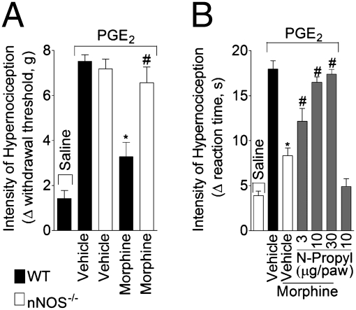
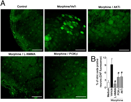
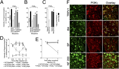
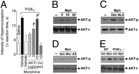
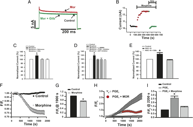
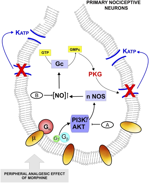
Morphine is one of the most prescribed and effective drugs used for the treatment of acute and chronic pain conditions. In addition to its central effects, morphine can also produce peripheral analgesia. However, the mechanisms underlying this peripheral action of morphine have not yet been fully elucidated. Here, we show that the peripheral antinociceptive effect of morphine is lost in neuronal nitric-oxide synthase null mice and that morphine induces the production of nitric oxide in primary nociceptive neurons. The activation of the nitric-oxide pathway by morphine was dependent on an initial stimulation of PI3Kgamma/AKT protein kinase B (AKT) and culminated in increased activation of K(ATP) channels. In the latter, this intracellular signaling pathway might cause a hyperpolarization of nociceptive neurons, and it is fundamental for the direct blockade of inflammatory pain by morphine. This understanding offers new targets for analgesic drug development.


Conflict of interest statement

The authors declare no conflict of interest.

Figures

Fig. 1.
Fig. 1.
Morphine activates the nNOS/NO antinociceptive pathway. (A) Mechanical hypernociception was induced by the injection of PGE2 (30 ng/paw) in the paws of mice. After 30 min, morphine (10 μg/paw) was administrated to wild-type or nNOS-deficient mice (nNOS−/−). Hypernociception was evaluated 1 hour after morphine injection using the electronic von Frey test (n = 6). *P < 0.05 compared with wild type. (B) Mechanical hypernociception in rats was induced by intraplantar injection of PGE2 (100 ng/paw). The antinociceptive effect of a local injection of morphine (6 μg/paw 2 hour after PGE2 injection) on PGE2-induced hypernociception was prevented by treatment with a selective inhibitor of nNOS (N-propyl-L-arginine; 3–30 μg/paw 30 min before morphine injection; n = 7). *, P < 0.05 compared with vehicle treatment. #, P < 0.05 compared with morphine treatment.
Fig. 2.
Fig. 2.
Morphine stimulated NO production in primary sensitive neurons (the role of PI3Kγ/AKT). (A) Representative images of DRG slices after ex vivo incubation with morphine (10 μM) in the presence or absence of L-NMMA (10 μM), AS605240 (PI3KγI; 100 nM), and AKT inhibitor (100 nM). DAF-FM fluorescence (green) indicates NO production. (Scale bars, 100 μm.) (B) Quantitative analysis of percentage of DRG neurons that increased their DAF-FM fluorescence intensity. *P < 0.05 compared with medium treatment. #P < 0.05 compared with morphine treatment.
Fig. 3.
Fig. 3.
PI3Kγ expression in primary nociceptive neurons and its requirement for the peripheral antinociceptive action of morphine. (A–C) Mechanical hypernociception in rats was induced by ipsilateral injection of PGE2 (100 ng/paw). The antinociceptive effect of morphine (6 μg/paw 2 hours after PGE2 injection) on PGE2-induced hypernociception was prevented by treatment with an inhibitor of PI3Ks (30 min before morphine injection) or (A) LY294002 (3–30 μg/paw; n = 6) or with selective inhibition of PI3Kγ by (B) AS605240 (10–90 μg/paw; n = 7) or (C) ODN antisense (AS) against PI3Kγ (50 μg/it intrathecal/day for 4 days; n = 5). *, P < 0.05 compared with vehicle treatment. #, P < 0.05 compared with morphine or mismatch (MS) treatment. (D) In mice, mechanical hypernociception was induced by the injection of PGE2 (30 ng/paw). After 30 min, morphine (indicated arrow; 10 μg/paw) was injected in the wild-type (WT) or PI3Kγ−/− mice (n = 10). (E) Mice received an intraplantar injection of CFA (10 μL/paw). After 4 hours, morphine (indicated arrow; 20 μg/paw) or saline (Sal) was injected in the WT or PI3Kγ−/− mice. *P < 0.05 compared with vehicle treatment. #, P < 0.05 compared with wild type treated with morphine. (F) Confocal images of typical examples of anti-PI3Kγ immunoreactivity in subpopulations of rat DRG neurons labeled using binding to IB4 or using antibodies to TRPV1, substance P (SP), and neurofilament (NF) 200. Arrows indicate double-labeled neurons. (Scale bars, 50 μm.)
Fig. 4.
Fig. 4.
Participation of AKT in the peripheral antinociceptive effect of morphine. (A) The antinociceptive effect of morphine on PGE2-induced hypernociception was prevented by the treatment of rat paw with AKT selective inhibitor IV (AKTi; n = 10). *, P < 0.05 compared with vehicle treatment. #, P < 0.05 compared with morphine treatment. (B) In vitro stimulation of DRG primary culture neurons from rats with morphine (10 μM) increased the phosphorylation of AKT analyzed by Western blot. (C) Naloxone (NLX- 1 μM) preincubation (10 min) prevented AKT phosphorylation induced by morphine. (D) Preincubation with wortmannin (100 nM) and AS605240 (100 nM) also reduced morphine-induced AKT phosphorylation. (E) Incubation of DRG-cultured neurons from WT mice with morphine also increased AKT phosphorylation but not in neurons from PI3K−/− mice.
Fig. 5.
Fig. 5.
Morphine increases KATP channel currents in primary nociceptive neurons (the role of the PI3Kγ/AKT/NO pathway). (A and B) Under voltage-clamp conditions, incubation of DRG neurons with morphine (10 μM, red line) elicited sustained increases in total whole-cell K+ currents that were not observed in the presence of glibenclamide (10 μM, green line). (C and D) The effect of morphine was not observed when the cells were incubated with naloxone (1 μM), L-NMMA (10 μM), PI3Kγ selective inhibitor (AS605240; 100 nM), and AKT inhibitor (100 nM). (E) In the same conditions, DRG neurons were incubated with NO donor (NOC-18; 10 μM) in the absence or presence of glibenclamide (10 μM). (F) Morphine causes a hyperpolarization of primary nociceptive neurons. Basal fluorescence intensity [DiBAC4(3)] was monitored during 5 min (only the last 100 s are present) followed by the incubation with morphine (10 μM) for 20 min. (G) Analyses of the maximal changes taken at 1,900 s in the membrane potential caused by morphine (control n = 32; morphine n = 9) are shown. (H and I) Morphine attenuates depolarization of nociceptive neurons caused by PGE2. Basal fluorescence of neurons was measured followed by incubation with PGE2 (1 μM; n = 26). After 15 min, morphine (10 μM) was added, and the fluorescence was monitored for 20 min (n = 24). *, P < 0.05 compared with medium treatment. #, P < 0.05 compared with morphine or DETA-NONOate (NOC-18) NOC-18 treatment.
Fig. 6.
Fig. 6.
Schematic representation of the molecular basis of morphine peripheral analgesia. The activation of opioid receptors in primary nociceptive neurons by morphine triggers the activation of the PI3Kγ/AKT pathway that in turn might cause the stimulation of nNOS and an increase in NO production. In last instance, NO, indirectly through stimulation of cGMP/PKG, causes the up-regulation of KATP currents and promotes the hyperpolarization of primary nociceptive neurons. The results described in the figure indicate the following treatments: (A) wortmannin, AS605240, LY294002, and AKT inhibitor IV, and (B) N-propyl-L-arginine. Morphine did not showed peripheral antinociceptive effect in (B) nNOS−/− and (A) PI3Kγ−/− mice.

References

    1. Ferreira SH, Nakamura M. II—Prostaglandin hyperalgesia: The peripheral analgesic activity of morphine, enkephalins and opioid antagonists. Prostaglandins. 1979;18:191–200. - PubMed
    1. Ferreira SH, Nakamura M. I—Prostaglandin hyperalgesia, a cAMP/Ca2+ dependent process. Prostaglandins. 1979;18:179–190. - PubMed
    1. Collier HO, Roy AC. Morphine-like drugs inhibit the stimulation of E prostaglandins of cyclic AMP formation by rat brain homogenate. Nature. 1974;248:24–27. - PubMed
    1. Levine JD, Taiwo YO. Involvement of the mu-opiate receptor in peripheral analgesia. Neuroscience. 1989;32:571–575. - PubMed
    1. Vetter I, Wyse BD, Monteith GR, Roberts-Thomson SJ, Cabot PJ. The mu opioid agonist morphine modulates potentiation of capsaicin-evoked TRPV1 responses through a cyclic AMP-dependent protein kinase A pathway. Mol Pain. 2006;2:22. - PMC - PubMed

Publication types

MeSH terms

LinkOut - more resources