. 2010 Mar 2;107(9):4135-40.
doi: 10.1073/pnas.0910632107. Epub 2010 Feb 10.

Electrostatics of aquaporin and aquaglyceroporin channels correlates with their transport selectivity

Affiliations

Electrostatics of aquaporin and aquaglyceroporin channels correlates with their transport selectivity

Romina Oliva et al. Proc Natl Acad Sci U S A. .

Abstract

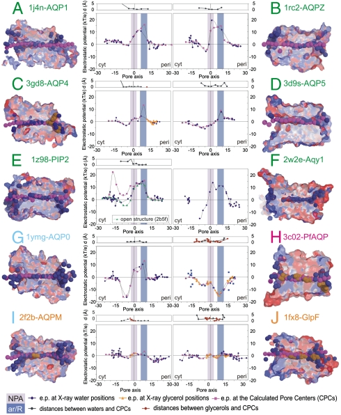
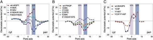
Aquaporins are homotetrameric channel proteins, which allow the diffusion of water and small solutes across biological membranes. According to their transport function, aquaporins can be divided into "orthodox aquaporins", which allow the flux of water molecules only, and "aquaglyceroporins", which facilitate the diffusion of glycerol and other small solutes in addition to water. The contribution of individual residues in the pore to the selectivity of orthodox aquaporins and aquaglyceroporins is not yet fully understood. To gain insights into aquaporin selectivity, we focused on the sequence variation and electrostatics of their channels. The continuum Poisson-Boltzmann electrostatic potential along the channel was calculated and compared for ten three-dimensional-structures which are representatives of different aquaporin subfamilies, and a panel of functionally characterized mutants, for which high-accuracy three-dimensional-models could be derived. Interestingly, specific electrostatic profiles associated with the main selectivity to water or glycerol could be identified. In particular: (i) orthodox aquaporins showed a distinctive electrostatic potential maximum at the periplasmic side of the channel around the aromatic/Arg (ar/R) constriction site; (ii) aquaporin-0 (AQP0), a mammalian aquaporin with considerably low water permeability, had an additional deep minimum at the cytoplasmic side; (iii) aquaglyceroporins showed a rather flat potential all along the channel; and (iv) the bifunctional protozoan PfAQP had an unusual all negative profile. Evaluation of electrostatics of the mutants, along with a thorough sequence analysis of the aquaporin pore-lining residues, illuminated the contribution of specific residues to the electrostatics of the channels and possibly to their selectivity.


Conflict of interest statement

The authors declare no conflict of interest.

Figures

Fig. 1.
Fig. 1.
Electrostatic potentials into the channels, calculated at the crystallographic positions of water/glycerol molecules and at the pore centers predicted by the PoreWalker program (33). Orthodox aquaporins are labeled in green, the low water conductance AQP0 in cyan, aquaglyceroporins in orange, and the bifunctional PfAQP in magenta. Sections of the corresponding PDB structures are shown colored according to the electrostatic potentials. X-ray water and glycerol atoms inside the channels are shown as deep blue and orange surfaces, respectively. Dummy atoms at the geometrical centers of the pores are shown as magenta surfaces.
Fig. 2.
Fig. 2.
Electrostatic potentials calculated at pore centers predicted by PoreWalker (33) for: A) AQP1 mutants, B) PfAQP mutants, and C) AQP0 mutants.
Fig. 3.
Fig. 3.
Representation of sequence and structural features of the pores. In each quadrant, the pore is orientated from the intracellular to the extracellular side and is shown in its sequence (bottom) and three-dimensional-structure (top). Acidic residues are colored in red, basic residues in blue and polar residues in purple. Three-dimensional-structure representation (top): for each structure, all the residues lining the pore are shown and residues mutated and/or discussed in the text are labeled. Sequence representation (bottom): corresponding pore-lining residues are shown as sequence logos. Note that logo numbers correspond to the structural position of the residues along the channel axis and not to their sequence position. For details on this representation of the pore sequence see (39). The logo positions 9–44 line the inner region of the channel. The two asparagines of the NPA motifs are found at the logo positions 23 and 28. Arg of the ar/R selectivity filter occupies the logo position 30, while the other residues in the ar/R filter are located at the logo positions 32, 35, and 36.

Similar articles

Cited by

References

    1. Wu B, Beitz E. Aquaporins with selectivity for unconventional permeants. Cell Mol Life Sci. 2007;64:2413–2421. - PMC - PubMed
    1. Zardoya R. Phylogeny and evolution of the major intrinsic protein family. Biol Cell. 2005;97:397–414. - PubMed
    1. Quigley F, Rosenberg JM, Shachar-Hill Y, Bohnert HJ. From genome to function: The Arabidopsis aquaporins. Genome Biol. 2002;3 RESEARCH0001. - PMC - PubMed
    1. Magni F, et al. Proteomic knowledge of human aquaporins. Proteomics. 2006;6:5637–5649. - PubMed
    1. Fischer G, et al. Crystal structure of a yeast aquaporin at 1.15 angstrom reveals a novel gating mechanism. PLoS Biol. 2009;7:e1000130. - PMC - PubMed

Publication types

LinkOut - more resources