. 2010 Mar 15;184(6):2814-24.
doi: 10.4049/jimmunol.0902125. Epub 2010 Feb 10.

Neisseria gonorrhoeae enhances HIV-1 infection of primary resting CD4+ T cells through TLR2 activation

Affiliations

Neisseria gonorrhoeae enhances HIV-1 infection of primary resting CD4+ T cells through TLR2 activation

Jian Ding et al. J Immunol. .

Abstract

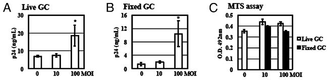
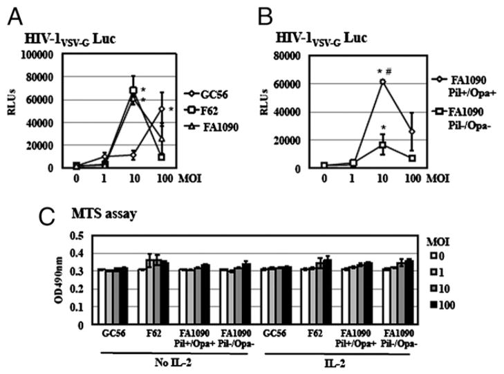
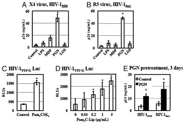
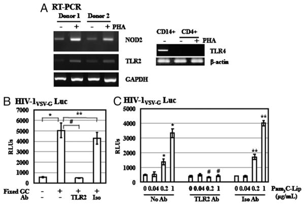
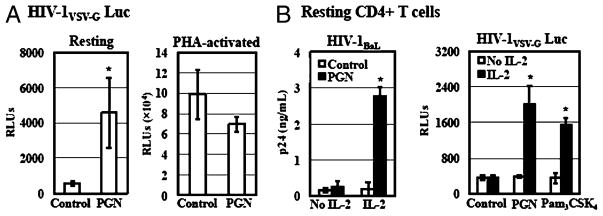
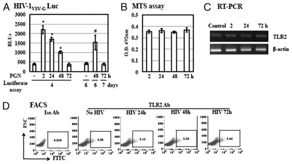
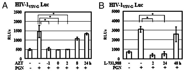
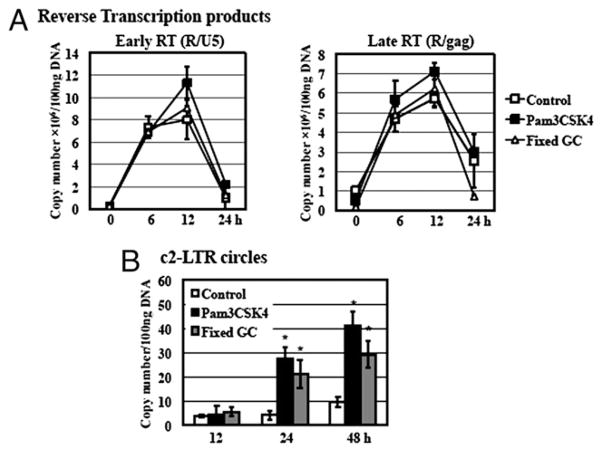
Sexually transmitted infections increase the likelihood of HIV-1 transmission. We investigated the effect of Neisseria gonorrheae (gonococcus [GC]) exposure on HIV replication in primary resting CD4(+) T cells, a major HIV target cell during the early stage of sexual transmission of HIV. GC and TLR2 agonists, such as peptidylglycan (PGN), Pam(3)CSK(4), and Pam(3)C-Lip, a GC-derived synthetic lipopeptide, but not TLR4 agonists including LPS or GC lipooligosaccharide enhanced HIV-1 infection of primary resting CD4(+) T cells after viral entry. Pretreatment of CD4(+) cells with PGN also promoted HIV infection. Anti-TLR2 Abs abolished the HIV enhancing effect of GC and Pam(3)C-Lip, indicating that GC-mediated enhancement of HIV infection of resting CD4(+) T cells was through TLR2. IL-2 was required for TLR2-mediated HIV enhancement. PGN and GC induced cell surface expression of T cell activation markers and HIV coreceptors, CCR5 and CXCR4. The maximal postentry HIV enhancing effect was achieved when PGN was added immediately after viral exposure. Kinetic studies and analysis of HIV DNA products indicated that GC exposure and TLR2 activation enhanced HIV infection at the step of nuclear import. We conclude that GC enhanced HIV infection of primary resting CD4(+) T cells through TLR2 activation, which both increased the susceptibility of primary CD4(+) T cells to HIV infection as well as enhanced HIV-infected CD4(+) T cells at the early stage of HIV life cycle after entry. This study provides a molecular mechanism by which nonulcerative sexually transmitted infections mediate enhancement of HIV infection and has implication for HIV prevention and therapeutics.


Conflict of interest statement

Disclosures

The authors have no financial conflicts of interest.

Figures

FIGURE 1
FIGURE 1
GC exposure enhances HIV-1 infection of primary resting CD4+ T cells. Primary resting CD4+ T cells were infected with HIV-1BaL at an MOI of 0.05 at 37°C for 2 h. After washing off unbound virus, infected cells were exposed to live (A) or fixed (B) GC (ATCC strain 43069) at various MOIs in the presence of IL-2. HIV production was determined by measuring the level of HIV capsid protein p24 at day 14 after viral infection using HIV p24 ELISA. C, Primary resting CD4+ T cells (1 × 10⁴ cells per well) were exposed to live GC or fixed GC at 37°C for 72 h in the presence of IL-2. Cell proliferation was examined by the MTS assay (Promega CellTiter96 aqueous one solution cell proliferation assay). Data are means ± SD of triplicate sample. Difference in HIV infection between samples without GC exposure and GC-exposed samples at an MOI of 100 is significant as calculated by the two-tailed, paired Student t test. *p < 0.05. There is no difference in cell proliferation between samples with or without GC exposure (p > 0.05). Data represent three independent experiments.
FIGURE 2
FIGURE 2
Multiple GC strains enhanced HIV-1 infection of primary resting CD4+ T cells. A, Primary resting CD4+ T cells were infected with pseudotyped HIV-1VSV-G luciferase reporter virus at 37°C for 2 h. After washing off unbound virus, infected cells were exposed to various strains of fixed GC in the presence of IL-2 for 4 d before measuring luciferase activity. There is a significant difference between samples with and without GC-exposure. *p < 0.05. B, Resting CD4+ T cells were infected with pseudotyped HIV-1VSV-G luciferase reporter virus at 37°C for 2 h, washed, and then treated with FA1090 Pil+/Opa+ or FA1090 Pil/Opa in the presence of IL-2. Luciferase activity was measured at 4 d postinfection. The difference between cells treated with FA1090 Pil+/Opa+ and FA1090 Pil/Opa is significant. #p < 0.05. C, Resting CD4+ T cells (1 × 10⁴ cells per well) were exposed to various strains of fixed GC at MOIs of 1, 10, and 100 at 37°C for 72 h in the presence of IL-2. Cell proliferation was examined by the MTS assay. Data are means ± SD of triplicate sample. Data represent two independent experiments.
FIGURE 3
FIGURE 3
TLR2 activation promotes HIV replication in primary resting CD4+ T cells. A and B, Primary resting CD4+ T cells were infected with X4 HIV-1IIIB or R5 HIV-1BaL at an MOI of 0.05 at 37°C for 2 h. After washing off unbound virus, infected cells were then treated with LPS (1 μg/ml), MDP (20 μg/ml), PGN (20 μg/ml), or GC DOV LOS (0.1 μg/ml) in the presence of IL-2 at 37°C. Infected cells without treatment were included as a control. The p24 level in the culture supernatant was determined on day 7 postinfection. C, Resting CD4+ T cells were infected with pseudotyped HIV-1VSV-G luciferase reporter virus at 37°C for 2 h and then treated with Pam3CSK4 at 5 μg/ml with IL-2 for 4 d before measuring luciferase assay. D, Resting CD4+ T cells were infected with pseudotyped HIV-1VSV-G luciferase reporter virus and then exposed to Pam3C-Lip at various concentrations with IL-2 for 4 d before measuring luciferase assay. E, Resting CD4+ T cells were treated with PGN (20 μg/ml) for 3 d, followed by washing. PGN-treated primary CD4+ cells were infected with HIV-1IIIB or HIV-1BaL. The p24 level in the culture supernatant was determined on day 7 postinfection. Data are means ±SD of triplicate sample and represent seven independent experiments. The difference in HIV infection between samples with and without TLR2 agonists treatment is significant as calculated by the two-tailed, paired Student t test. *p < 0.05.
FIGURE 4
FIGURE 4
Enhancement of HIV infection of primary resting CD4+ T cells by GC is mediated through TLR2 signaling pathway. A, Gene expression of NOD2, TLR2, and TLR4 in resting CD4+ T cells and PHA-activated CD4+ T cells was determined by RT-PCR analysis. Total RNA from CD14+ cells was included as a control for analysis of TLR4 gene expression. B, To determine the role of TLR2 in GC-mediated enhancement of HIV infection, resting CD4+ T cells were infected with pseudotyped HIV-1VSV-G luciferase reporter virus at 37°C for 2 h. After washing off unbound virus, infected cells were treated with anti-TLR2 Ab or isotype control Ab at 10 μg/ml for 1 h. Cells were exposed to fixed GC (ATCC strain 43069) at an MOI of 50 with IL-2 and cultured for 4 d before measurement of luciferase activity. C, Resting CD4+ T cells were infected with pseudotyped HIV-1VSV-G luciferase reporter virus and treated with anti-TLR2 Ab or isotype control Ab as described previously. Cells were then exposed to Pam3C-Lip at various concentrations in the presence of IL-2 for 4 d. Data are means ±SD of triplicate sample and represent two independent experiments. There is a significant difference between samples with and without GC or Pam3C-Lip treatment (*p < 0.05) as well as between stimulated cells in the presence or absence of anti-TLR2 Ab (#p < 0.05). There is no difference between stimulated samples in the presence or absence of isotype control Ab (**p < 0.05).
FIGURE 5
FIGURE 5
TLR2-mediated HIV enhancement occurs in resting but not PHA-activated CD4+ T cells and IL-2 is required for the HIV enhancing effect. A, Resting CD4+ T cells were activated with or without PHA and IL-2 for 3 d. Resting CD4+ T cells with or without PHA activation were infected with pseudotyped HIV-1VSV-G luciferase reporter virus for 2 h. Infected cells were then treated with PGN at 20 μg/ml in the presence of IL-2 and cultured for 4 d before measurement of luciferase activity. B, Resting CD4+ T cells were infected with HIV-1BaL at an MOI of 0.05 or pseudotyped HIV-1VSV-G luciferase reporter virus. Infected cells were treated with Pam3CSK4 at 5μg/ml or PGN at 20 μg/ml in the presence or absence of IL-2. HIV-infected cells without treatment were included as a control. Viral production by primary CD4+ T cells infected with HIV-1BaL was determined by measuring the p24 level in the culture supernatant at day 7 postinfection. Luciferase activity was determined at day 4 postinfection for the single-cycle infection assay. Data are means ±SD of triplicate sample and represent three independent experiments. *p < 0.05.
FIGURE 6
FIGURE 6
The maximal HIV enhancing effect of the TLR2 activation is achieved when PGN is added at an early stage of the HIV life cycle. A, Resting CD4+ T cells were infected with pseudotyped HIV-1VSV-G luciferase reporter virus for 2 h. Infected cells were cultured with IL-2 and then treated with PGN at 20 μg/ml at 2, 24, 48, or 72 h postinfection before measurement of luciferase activity at day 4, 6, or 7 postinfection. There is a significant difference between samples without PGN and with PGN at 2, 24, or 48 h postinfection (*p < 0.05). When samples were harvested at day 6 postinfection the difference between cells with PGN at 48 h postinfection and without treatment is significant (#p < 0.05). B, The viability of resting CD4+ T cells postinfection with pseudotyped HIV-1VSV-G luciferase reporter virus was assessed by MTS assay at 2, 24, 48, and 72 h after viral infection. C, Total RNA from resting CD4+ T cells with infection by pseudotyped HIV-1VSV-G reporter virus for 2, 24, or 72 h was prepared. TLR2 gene expression of TLR2 was analyzed by RT-PCR. Samples from mock-infected resting CD4+ T cells were also included as a control. D, Cell surface expression of TLR2 on HIV-infected resting CD4+ T cells was determined by FACS analysis. Data are means ±SD of triplicate sample and represent three independent experiments.
FIGURE 7
FIGURE 7
The kinetics of HIV infection in TLR2-activated resting CD4+ T cells in the presence of inhibitors for reverse transcriptase and integrase. A, Resting CD4+ T cells were infected with pseudotyped HIV-VSV-G luciferase reporter virus for 2 h. Infected cells were treated with AZT at 5 μM at 1 h before infection (−1 h), at the same time during HIV exposure (0 h) or at 2, 8, and 24 h postinfection. PGN at 20 μg/ml was added at 2 h after viral infection. Infected cells were cultured in the presence of IL-2 for 4 d before measurement of luciferase activity. B, In a single-cycle infection assay, resting CD4+ T cells were infected with pseudotyped HIV-1 reporter virus for 2 h, treated with PGN at 20 μg/ml, and cultured in complete media with IL-2. L-731,988 at 10 μM was added to cell culture at 2, 24, and 48 h postinfection. Luciferase activity was measured on day 4 postinfection. Data are means ± SD of triplicate sample and represent three independent experiments.
FIGURE 8
FIGURE 8
TLR2 activation and GC exposure promote HIV infection at the step of nuclear import. Resting CD4+ T cells were infected with pseudotyped HIV-1VSV-G luciferase reporter virus and then treated with Pam3CSK4 at 5 μg/ml or fixed GC (ATCC strain 43069) at an MOI of 50 in the presence of IL-2. DNA was prepared from TLR2-activated or GC-exposed HIV-infected cells at 0, 6, 12, 24, and 48 h postinfection. DNA from mock-infected primary resting CD4+ T cells was also isolated as a control. A, Quantitation of HIV-1 early strong-stop (R/U5) and late full-length (R/gag) RT products was performed by quantitative real-time PCR analysis. Data are means ± SD of triplicate sample and represent three independent experiments. Differences in the levels of early and late RT products between samples with or without exposure to GC or TLR2 agonists are not significant as calculated by the two-tailed, paired Student t test, p < 0.05. B, To assess whether TLR2 activation or GC exposure affected HIV-1 nuclear import, quantitative real-time PCR analysis was performed to measure c2-LTR circles in infected CD4+ T cells with or without Pam3CSK4 or fixed GC treatment for 12, 24, and 48 h. Plasmid pTA2LTR at different copy numbers was used to generate a standard for c2-LTR circles detection. The detection limit of the PCR analysis was 10 copies. There is a significant difference between samples with or without exposure to TLR2 agonists or GC (*p < 0.05) at 24 and 48 h postinfection.

Similar articles

Cited by

References

    1. Hall HI, Song R, Rhodes P, Prejean J, An Q, Lee LM, Karon J, Brookmeyer R, Kaplan EH, McKenna MT, et al. Estimation of HIV incidence in the United States. JAMA. 2008;300:520–529. - PMC - PubMed
    1. Cohen MS, Miller WC. Sexually transmitted diseases and human immunodeficiency virus infection: cause, effect, or both? Int J Infect Dis. 1998;3:1–4. - PubMed
    1. Plummer FA, Simonsen JN, Cameron DW, Ndinya-Achola JO, Kreiss JK, Gakinya MN, Waiyaki P, Cheang M, Piot P, Ronald AR, et al. Cofactors in male-female sexual transmission of human immunodeficiency virus type 1. J Infect Dis. 1991;163:233–239. - PubMed
    1. Cameron DW, Simonsen JN, D’Costa LJ, Ronald AR, Maitha GM, Gakinya MN, Cheang M, Ndinya-Achola JO, Piot P, Brunham RC, et al. Female to male transmission of human immunodeficiency virus type 1: risk factors for seroconversion in men. Lancet. 1989;2:403–407. - PubMed
    1. Ghys PD, Fransen K, Diallo MO, Ettiègne-Traoré V, Coulibaly IM, Yeboué KM, Kalish ML, Maurice C, Whitaker JP, Greenberg AE, Laga M. The associations between cervicovaginal HIV shedding, sexually transmitted diseases and immunosuppression in female sex workers in Abidjan, Côte d’Ivoire. AIDS. 1997;11:F85–F93. - PubMed

Publication types

MeSH terms