

Gray matter perfusion correlates with disease severity in ALS
- PMID: 20147656
- PMCID: PMC2839193
- DOI: 10.1212/WNL.0b013e3181d3e2dd
Gray matter perfusion correlates with disease severity in ALS
Abstract
Objective: The goal of this study is to determine if regional brain perfusion, as measured by arterial spin labeling (ASL) MRI, is correlated with clinical measures of amyotrophic lateral sclerosis (ALS) disease severity. The presence of such a relationship would indicate a possible role for ASL perfusion as a marker of disease severity and upper motor neuron involvement in ALS.
Methods: Disease severity was assessed in 16 subjects with ALS (age 54 +/- 11) using the Amyotrophic Lateral Sclerosis Functional Rating Scale (ALSFRS) and the pulmonary function measure, forced vital capacity (FVC). Upper motor neuron involvement was assessed by testing rapid tapping of the fingers and feet. Magnetic resonance perfusion images were coregistered with structural T1-weighted MRI, corrected for partial volume effects using the structural images and normalized to a study-specific atlas. Correlations between perfusion and ALS disease severity were analyzed, using statistical parametric mapping, and including age as a factor. Analyses were adjusted for multiple clusters.
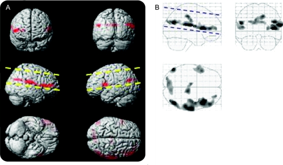
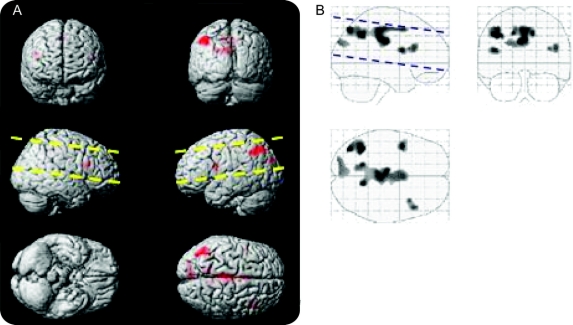
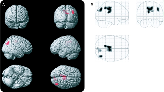
Result: ALS severity, as measured by the ALSFRS and FVC, was correlated with gray matter perfusion. This correlation was predominantly observed in the hemisphere contralateral to the more affected limbs. ALSFRS scores correlated with perfusion in the contralateral frontal and parietal lobe (p < 0.001) and ipsilateral frontal lobe (p < 0.02). FVC scores correlated with gray matter perfusion in contralateral frontal lobe (p < 0.001). Upper motor neuron involvement, as measured by rapid finger tapping, correlated bilaterally with perfusion in the middle cingulate gyrus (p < 0.001).
Conclusion: Amyotrophic lateral sclerosis (ALS) severity is correlated with brain perfusion as measured by arterial spin labeling (ASL) perfusion. This correlation appears to be independent of brain atrophy. ASL perfusion may be a useful tool for monitoring disease progression and assessing treatment effects in ALS.
Figures
References
-
- Kassubek J, Unrath A, Huppertz HJ, et al. Global brain atrophy and corticospinal tract alterations in ALS, as investigated by voxel-based morphometry of 3-D MRI. Amyotroph Lateral Scler Other Motor Neuron Disord 2005;6:213–220. - PubMed
-
- Ellis CM, Simmons A, Jones DK, et al. Diffusion tensor MRI assesses corticospinal tract damage in ALS. Neurology 1999;53:1051–1058. - PubMed
-
- Abe K, Takanashi M, Watanabe Y, et al. Decrease in N-acetylaspartate/creatine ratio in the motor area and the frontal lobe in amyotrophic lateral sclerosis. Neuroradiology 2001;43:537–541. - PubMed
-
- Beall DP, Martin D, Chin BB. Decreased bilateral frontal lobe perfusion in dementia of amyotrophic lateral sclerosis. Clin Nucl Med 1998;23:855–856. - PubMed
Publication types
MeSH terms
Grants and funding
- P50 AG023501/AG/NIA NIH HHS/United States
- P41 RR 023953/RR/NCRR NIH HHS/United States
- R01 AG10897/AG/NIA NIH HHS/United States
- R01 NS 44887/NS/NINDS NIH HHS/United States
- P01AG012435/AG/NIA NIH HHS/United States
- P50AG23501/AG/NIA NIH HHS/United States
- P01AG19724/AG/NIA NIH HHS/United States
- R01AG032306/AG/NIA NIH HHS/United States
- K01 HL73152/HL/NHLBI NIH HHS/United States
- U01AG024904/AG/NIA NIH HHS/United States
- P41 RR023953/RR/NCRR NIH HHS/United States
- R24 RR021992/RR/NCRR NIH HHS/United States
- R01NS031966/NS/NINDS NIH HHS/United States
LinkOut - more resources
Full Text Sources
Medical
Miscellaneous