Review
. 2010 Apr 9;285(15):11061-7.
doi: 10.1074/jbc.R109.072181. Epub 2010 Feb 10.

Chemical inducers of autophagy that enhance the clearance of mutant proteins in neurodegenerative diseases

Affiliations
Review

Chemical inducers of autophagy that enhance the clearance of mutant proteins in neurodegenerative diseases

Maurizio Renna et al. J Biol Chem. .

Abstract

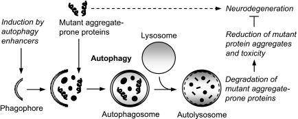
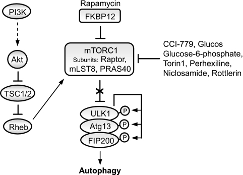
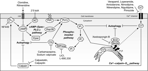
Many of the neurodegenerative diseases that afflict people are caused by intracytoplasmic aggregate-prone proteins. These include Parkinson disease, tauopathies, and polyglutamine expansion diseases such as Huntington disease. In Mendelian forms of these diseases, the mutations generally confer toxic novel functions on the relevant proteins. Thus, one potential strategy for dealing with these mutant proteins is to enhance their degradation. This can be achieved by up-regulating macroautophagy, which we will henceforth call autophagy. In this minireview, we will consider the reasons why autophagy up-regulation may be a powerful strategy for these diseases. In addition, we will consider some of the drugs and associated signaling pathways that can be used to induce autophagy with these therapeutic aims in mind.


Figures

FIGURE 1.
FIGURE 1.
Autophagy as a protective pathway for neurodegenerative diseases. Autophagy is a major degradation pathway for the clearance of various intracytosolic toxic aggregate-prone proteins associated with neurodegenerative diseases. Chemical induction of autophagy by autophagy enhancers triggers cellular signaling pathways, leading to formation of double-membrane cytoplasmic structures called phagophores. These structures elongate and engulf mutant aggregate-prone proteins along with portions of the cytoplasm to form autophagosomes. Autophagosomes then ultimately fuse with the lysosomes to form autolysosomes, where their contents are degraded by acidic lysosomal hydrolases. Enhancing autophagic clearance of these mutant aggregate-prone proteins results in reduction of mutant protein aggregates and toxicity, which is protective in several models of neurodegenerative diseases.
FIGURE 2.
FIGURE 2.
Regulation of autophagy by the mTOR pathway. Autophagy is negatively regulated by mTOR, which is downstream in the phosphatidylinositol 3-kinase (PI3K) pathway. A diverse range of signals, such as growth factors and amino acids, regulates mTORC1 by inhibiting TSC1/2, thereby alleviating the inhibitory effect of TSC1/2 on Rheb, which subsequently activates mTORC1. Several kinases, such as Akt, signal to mTORC1 by phosphorylating TSC2 and inhibiting the activity of the TSC1/2 heterodimer. Rapamycin forms a complex with the immunophilin FKBP12, which inhibits the kinase activity of mTORC1. Inhibition of mTOR by rapamycin induces autophagy and enhances the clearance of mutant aggregate-prone proteins. The ULK1-Atg13-FIP200 complex acts as an integrator of the autophagy signals downstream of mTORC1. Under nutrient-rich conditions, mTORC1 suppresses autophagy by interacting with this complex and mediating phosphorylation-dependent inhibition of Atg13 and ULK1. Treatment with rapamycin dissociates mTOR from the complex, resulting in dephosphorylation-dependent activation of ULK1 and ULK1-mediated phosphorylations of Atg13, FIP200, and ULK1 itself, which triggers autophagy. Other chemical inhibitors of mTORC1 include CCI-779, glucose, glucose 6-phosphate, Torin1, perhexiline, niclosamide, and rottlerin. These may act directly or indirectly.
FIGURE 3.
FIGURE 3.
Cyclical mTOR-independent autophagy pathway with multiple drug targets for neurodegenerative diseases. Shown is a cyclical mTOR-independent pathway regulating mammalian autophagy, comprising cAMP-Epac-PLCϵ-IP3 and Ca2+-calpain-Gαs pathways, which has multiple drug targets for neurodegenerative diseases. Intracellular cAMP levels are increased by adenylyl cyclase (AC) activity, thus activating Epac, which then activates the small G-protein Rap2B, thereby activating PLCϵ. PLCϵ mediates the production of IP3 from phosphatidylinositol 4,5-bisphosphate (PIP2), thereby increasing the levels of IP3 that binds to ER-resident IP3Rs, leading to release of Ca2+ from the ER stores. Intracytosolic Ca2+ levels are also increased by L-type Ca2+ channel agonists. Elevated intracytosolic Ca2+ activates calpains, which then cleave and activate Gαs. In turn, activation of Gαs increases adenylyl cyclase activity to elevate cAMP levels, thereby forming a loop. Activation of this pathway inhibits autophagy. Multiple drug targets acting at distinct stages in this pathway trigger autophagy, such as imidazoline-1 receptor (I1R) agonists (clonidine and rilmenidine) and the adenylyl cyclase inhibitor 2′,5′-dideoxyadenosine (2′5′ddA), which decrease cAMP levels; agents that lower inositol (Ins) and IP3 levels (carbamazepine and sodium valproate); IMPase inhibitors that also reduce inositol and IP3 levels (lithium and L-690,330); Ca2+ channel blockers (verapamil, loperamide, amiodarone, nimodipine, nitrendipine, niguldipine, and pimozide); calpain inhibitors (calpastatin and calpeptin); and the Gαs inhibitor NF449. Furthermore, inhibition of the IP3R by xestospongin B also induces autophagy by disrupting the IP3R-beclin 1 complex and, consequently, the Bcl-2-beclin 1 autophagy inhibitory complex. Enhancing autophagy through this mTOR-independent pathway is protective in various models of HD.

References

    1. Rubinsztein D. C. (2006) Nature 443, 780–786 - PubMed
    1. Ross C. A., Poirier M. A. (2004) Nat. Med. 10, (suppl.) S10–S17 - PubMed
    1. Zoghbi H. Y., Orr H. T. (2000) Annu. Rev. Neurosci. 23, 217–247 - PubMed
    1. Imarisio S., Carmichael J., Korolchuk V., Chen C. W., Saiki S., Rose C., Krishna G., Davies J. E., Ttofi E., Underwood B. R., Rubinsztein D. C. (2008) Biochem. J. 412, 191–209 - PubMed
    1. Arrasate M., Mitra S., Schweitzer E. S., Segal M. R., Finkbeiner S. (2004) Nature 431, 805–810 - PubMed

Publication types