

Amygdala transcriptome and cellular mechanisms underlying stress-enhanced fear learning in a rat model of posttraumatic stress disorder
- PMID: 20147889
- PMCID: PMC3040562
- DOI: 10.1038/npp.2010.10
Amygdala transcriptome and cellular mechanisms underlying stress-enhanced fear learning in a rat model of posttraumatic stress disorder
Abstract
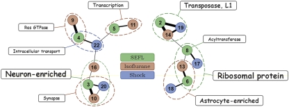
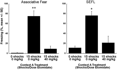
Severe stress or trauma can cause permanent changes in brain circuitry, leading to dysregulation of fear responses and the development of posttraumatic stress disorder (PTSD). To date, little is known about the molecular mechanisms underlying stress-induced long-term plasticity in fear circuits. We addressed this question by using global gene expression profiling in an animal model of PTSD, stress-enhanced fear learning (SEFL). A total of 15 footshocks were used to induce SEFL and the volatile anesthetic isoflurane was used to suppress the behavioral effects of stress. Gene expression in lateral/basolateral amygdala was measured using microarrays at 3 weeks after the exposure to different combinations of shock and isoflurane. Shock produced robust effects on amygdalar transcriptome and isoflurane blocked or reversed many of the stress-induced changes. We used a modular approach to molecular profiles of shock and isoflurane and built a network of regulated genes, functional categories, and cell types that represent a mechanistic foundation of perturbation-induced plasticity in the amygdala. This analysis partitioned perturbation-induced changes in gene expression into neuron- and astrocyte-specific changes, highlighting a previously underappreciated role of astroglia in amygdalar plasticity. Many neuron-enriched genes were highly correlated with astrocyte-enriched genes, suggesting coordinated transcriptional responses to environmental challenges in these cell types. Several individual genes were validated using RT-PCR and behavioral pharmacology. This study is the first to propose specific cellular and molecular mechanisms underlying SEFL, an animal model of PTSD, and to nominate novel molecular and cellular targets with potential for therapeutic intervention in PTSD, including glycine and neuropeptide systems, chromatin remodeling, and gliotransmission.
Figures


Similar articles
-
Isoflurane suppresses stress-enhanced fear learning in a rodent model of post-traumatic stress disorder.Anesthesiology. 2009 Mar;110(3):487-95. doi: 10.1097/ALN.0b013e3181974f3e. Anesthesiology. 2009. PMID: 19212264 Free PMC article.
-
Changes in Galanin Systems in a Rat Model of Post-Traumatic Stress Disorder (PTSD).PLoS One. 2016 Dec 1;11(12):e0167569. doi: 10.1371/journal.pone.0167569. eCollection 2016. PLoS One. 2016. PMID: 27907151 Free PMC article.
-
Individual susceptibility or resistance to posttraumatic stress disorder-like behaviours.Behav Brain Res. 2020 May 27;386:112591. doi: 10.1016/j.bbr.2020.112591. Epub 2020 Mar 16. Behav Brain Res. 2020. PMID: 32194190
-
Neurogenetic Approaches to Stress and Fear in Humans as Pathophysiological Mechanisms for Posttraumatic Stress Disorder.Biol Psychiatry. 2018 May 15;83(10):810-820. doi: 10.1016/j.biopsych.2017.12.015. Epub 2018 Jan 10. Biol Psychiatry. 2018. PMID: 29454655 Review.
-
Fear conditioning, synaptic plasticity and the amygdala: implications for posttraumatic stress disorder.Trends Neurosci. 2012 Jan;35(1):24-35. doi: 10.1016/j.tins.2011.06.007. Epub 2011 Jul 26. Trends Neurosci. 2012. PMID: 21798604 Free PMC article. Review.
Cited by
-
Expression profiling associates blood and brain glucocorticoid receptor signaling with trauma-related individual differences in both sexes.Proc Natl Acad Sci U S A. 2014 Sep 16;111(37):13529-34. doi: 10.1073/pnas.1401660111. Epub 2014 Aug 11. Proc Natl Acad Sci U S A. 2014. PMID: 25114262 Free PMC article.
-
Mass Spectrometry-Based Approaches to Understand the Molecular Basis of Memory.Front Chem. 2016 Oct 14;4:40. doi: 10.3389/fchem.2016.00040. eCollection 2016. Front Chem. 2016. PMID: 27790611 Free PMC article. Review.
-
The effect of stress and reward on encoding future fear memories.Behav Brain Res. 2022 Jan 24;417:113587. doi: 10.1016/j.bbr.2021.113587. Epub 2021 Sep 17. Behav Brain Res. 2022. PMID: 34543677 Free PMC article. Review.
-
Small RNA Pathways That Protect the Somatic Genome.Int J Mol Sci. 2017 Apr 26;18(5):912. doi: 10.3390/ijms18050912. Int J Mol Sci. 2017. PMID: 28445427 Free PMC article. Review.
-
A New Outlook on Mental Illnesses: Glial Involvement Beyond the Glue.Front Cell Neurosci. 2015 Dec 16;9:468. doi: 10.3389/fncel.2015.00468. eCollection 2015. Front Cell Neurosci. 2015. PMID: 26733803 Free PMC article. Review.
References
-
- Amat J, Matus-Amat P, Watkins LR, Maier SF. Escapable and inescapable stress differentially alter extracellular levels of 5-HT in the basolateral amygdala of the rat. Brain Res. 1998;812:113–120. - PubMed
-
- American Psychiatric Association 1994Diagnostic and Statistical Manual of Mental DisordersIV edn.American Psychiatric Press: Washington, DC
Publication types
MeSH terms
Substances
Grants and funding
LinkOut - more resources
Full Text Sources
Other Literature Sources
Medical
