. 2010 May;3(3):280-90.
doi: 10.1038/mi.2010.2. Epub 2010 Feb 10.

HIV-1 sexual transmission: early events of HIV-1 infection of human cervico-vaginal tissue in an optimized ex vivo model

Affiliations

HIV-1 sexual transmission: early events of HIV-1 infection of human cervico-vaginal tissue in an optimized ex vivo model

E Saba et al. Mucosal Immunol. 2010 May.

Abstract

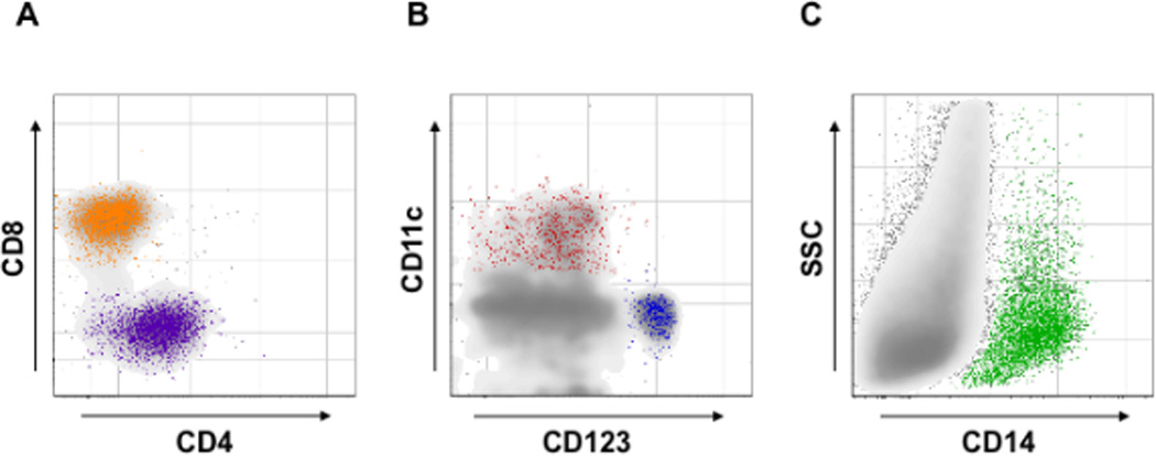
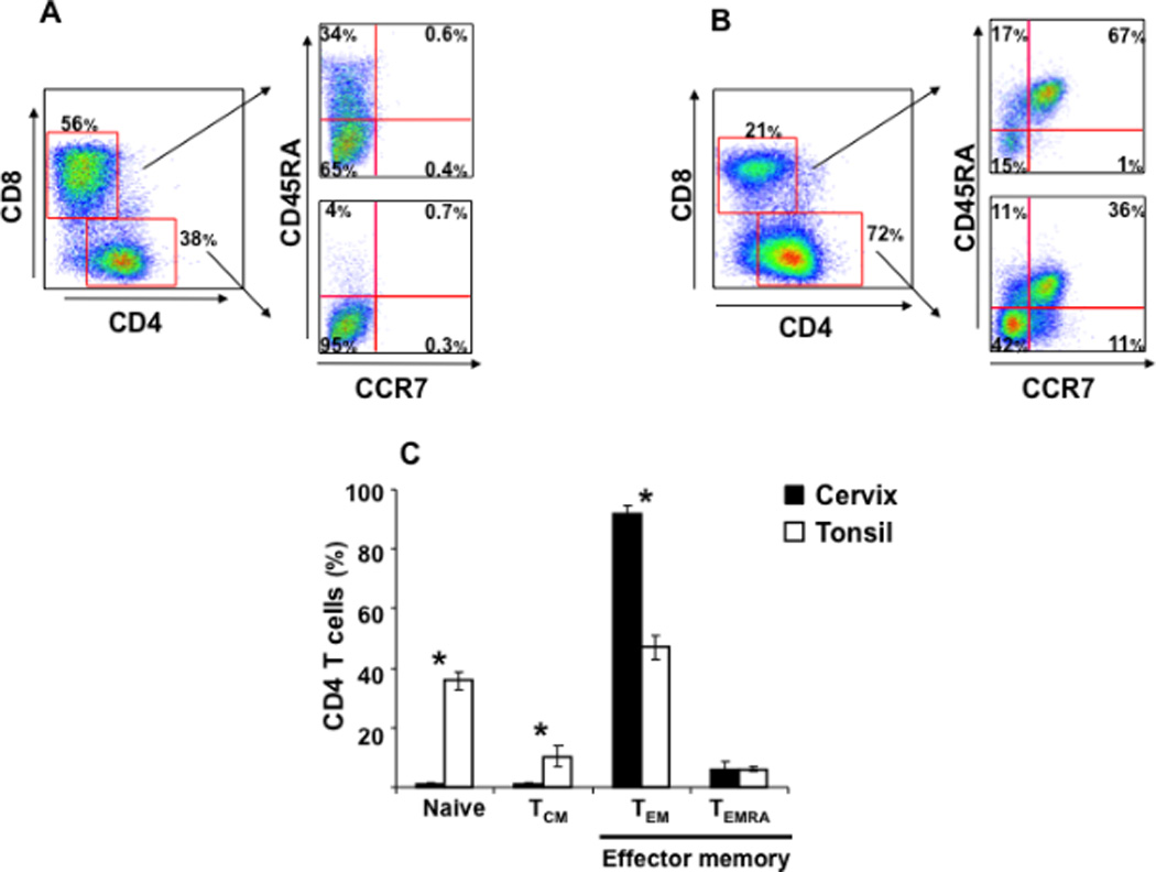
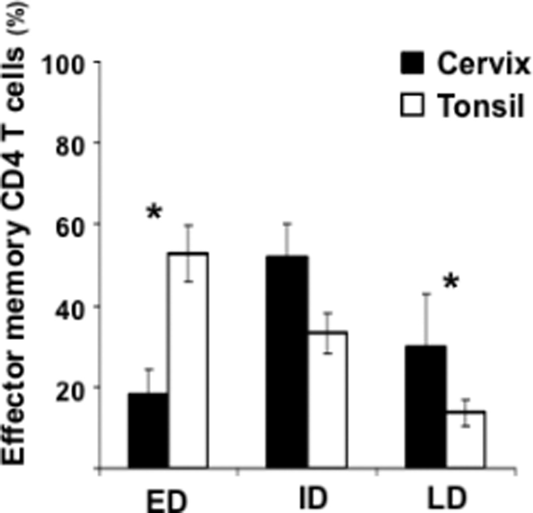
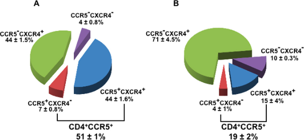
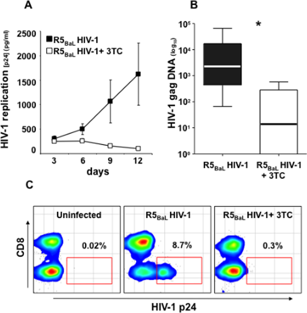
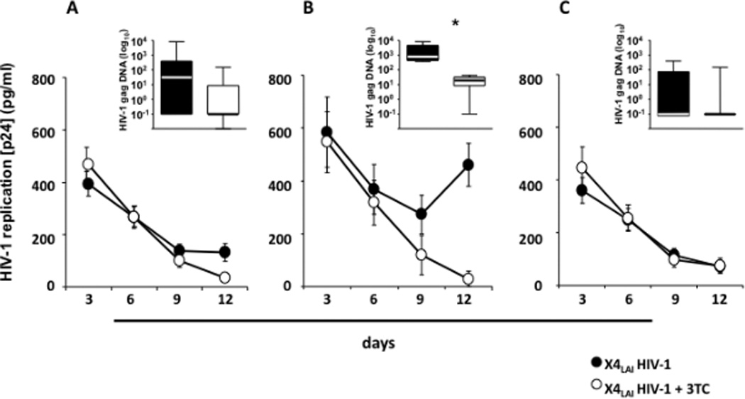
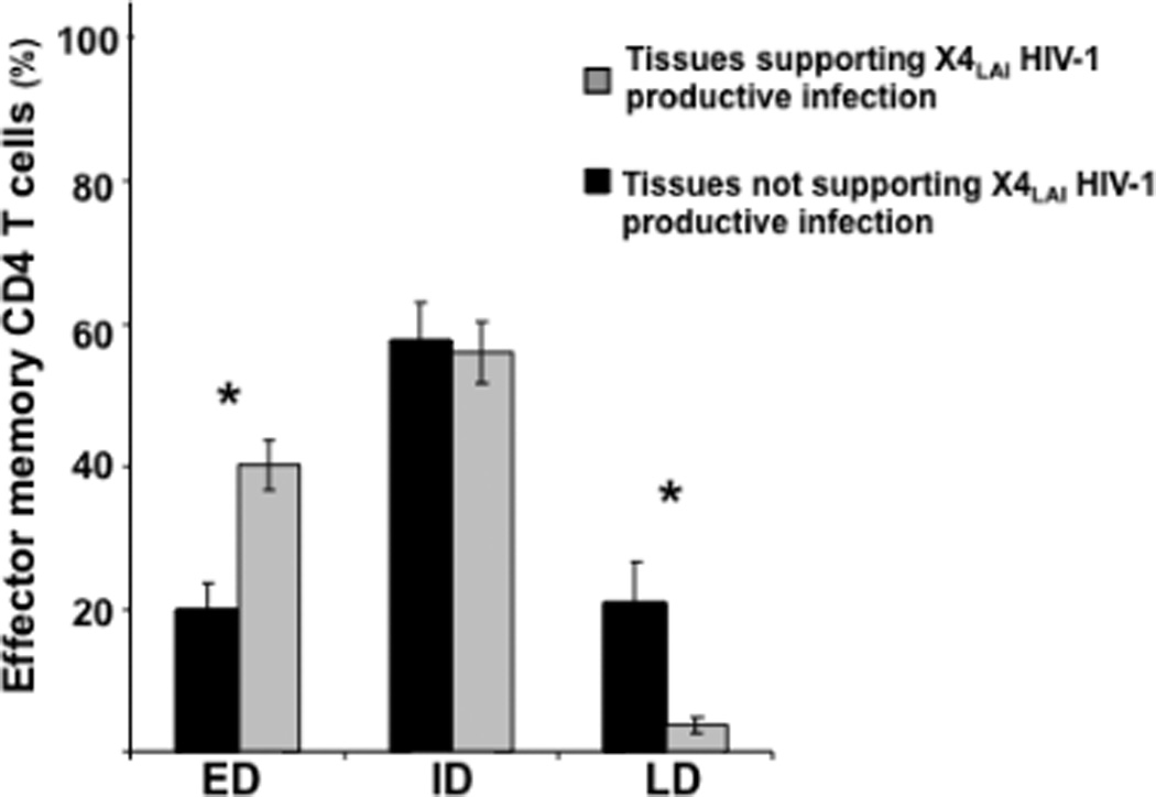
Infection and dissemination of human immunodeficiency virus (HIV)-1 through the female body after vaginal intercourse depends on the activation/differentiation status of mucosal CD4 T cells. In this study, we investigated this status and the susceptibility to HIV-1 infection of human cervico-vaginal tissue ex vivo. We found that virtually all T cells are of the effector memory phenotype with broad CC chemokine receptor 5 (CCR5) expression. As it does in vivo, human cervico-vaginal tissue ex vivo preferentially supports the productive infection of R5 HIV-1 rather than that of X4 HIV-1 in spite of the broad expression of CXC chemokine receptor 4 (CXCR4). X4 HIV-1 replicated only in the few tissues that were enriched in CD27(+)CD28(+) effector memory CD4 T cells. Productive infection of R5 HIV-1 occurred preferentially in activated CD38(+)CD4 T cells and was followed by a similar activation of HIV-1-uninfected (bystander) CD4 T cells that may amplify viral infection. These results provide new insights into the dependence of HIV-1 infection and dissemination on the activation/differentiation of cervico-vaginal lymphocytes.


Figures

Figure 1
Figure 1. Identification of T lymphocytes, dendritic cells and macrophages in human cervico-vaginal tissue
Cells suspension from digested fresh cervico-vaginal tissue blocks was stained for markers of T lymphocytes, dendritic cells [plasmacytoid (PDCs) and myeloid (MDCs)] and macrophages and analyzed with flow cytometry. Presented are bivariate dot plots of the distribution of CD4 and CD8 T cells (A), PDCs and MDCs (B), and macrophages (C). CD4- and CD8-positive T cells were identified gating on CD3+ lymphocytes. PDCs (CD123+) and MDCs (CD11c+) were identified gating on cells negative for CD3, CD14, CD16, CD56, CD19, CD20 and positive for HLA-DR. Macrophages (CD14+) were identified gating on CD3, CD56, CD19, CD20 negative cells. Presented plots are representative of experiments with cervico-vaginal tissues from five to twelve donors. Note that T lymphocytes, PDCs, MDCs, and macrophages that are involved in HIV-1 infection are present in human cervico-vaginal tissue.
Figure 2
Figure 2. T-lymphocyte subpopulations in human cervico-vaginal and tonsillar tissue
Cells suspension was stained for lymphocytic markers and analyzed with flow cytometry. Presented are bivariate dot plots of the distribution of different T-lymphocyte subpopulations for cervico-vaginal (A) and tonsillar (B) tissues. For each tissue, the distribution of CD4 and CD8 T cells is displayed in the left panel. The distribution of naïve, TCM, TEM, and TEMRA subpopulations among CD4 and CD8 T cells is displayed in the upper right panel and in the lower right panel, respectively. Presented plots are representative of experiments with cervico-vaginal tissues from eleven donors and tonsillar tissues from five donors. In C are presented the means ± SEM of the fractions of naïve, TCM, TEM, and TEMRA among CD4 T cells from these tissues. *Indicates a statistically significant difference with p<0.05. Note that the distribution of naïve, TCM, and TEM CD4 T cells in cervico-vaginal tissues is different from that in tonsillar tissues.
Figure 3
Figure 3. Distribution of early-, intermediate-. and late-differentiated effector memory CD4 T cells in human cervico-vaginal and tonsillar tissues
Early- (ED), intermediate- (ID) and late-differentiated (LD) effector memory CD4 T cells (TEM, TEMRA) were identified on the basis of the expression of CD27 and CD28 costimulatory molecules. Presented are means ± SEM of the fractions of ED, ID, and LD effector memory CD4 T cells in cervico-vaginal (n=10) and tonsillar (n=5) tissues. *Indicates a statistically significant difference with p<0.05. Note that the distribution of ED and LD effector memory CD4 T cells in cervico-vaginal tissues is different from that in tonsillar tissues.
Figure 4
Figure 4. Expression of HIV-1 coreceptors on CD4 T cells from human cervico-vaginal and tonsillar tissues
Cells isolated from fresh tissue blocks were stained for lymphocytic markers and analyzed with flow cytometry. Presented are means ± SEM of the fractions of total CD4 T cells expressing CCR5 and CXCR4 in cervico-vaginal (A, n=4) and tonsillar (B, n=3) tissues. Note that the fraction of CD4 T cells expressing CCR5 is higher in cervico-vaginal than in tonsillar tissue, while the fraction of CD4 T cells expressing CXCR4 is similar.
Figure 5
Figure 5. R5BaL HIV-1 infection of human cervico-vaginal tissues
Donor-matched blocks of human cervico-vaginal tissue were inoculated ex vivo with R5BaL HIV-1 and treated or not with 3TC (5 µM). A: Kinetics of release of p24gag by inoculated tissues. Presented are means ± SEM for tissues from 16 donors. For each donor, each datum point represents pooled viral release from 16 tissue blocks over 3-day periods. B: The amount of HIV-1 gag complete reverse transcripts at day 12 post-infection. Presented are medians and the interquartile ranges; the whiskers of the graph show the largest and smallest values. C: The distribution of HIV-1-infected cells in donor-matched tissue blocks: uninfected, inoculated with R5BaL HIV-1, inoculated with R5BAL, and treated with 3TC (5 µM). The amounts of HIV-1 p24gag-positive cells are expressed as percentage of total CD3+ T cells. Presented are bivariate density plots of a representative experiment with cervico-vaginal tissues from one out of 13 donors. *Indicates a statistically significant difference with p<0.05. Note that p24gag accumulation in culture medium measured by cytometric bead assay, production of HIV-1 complete reverse transcripts measured by real-time PCR, and identification of HIV-1-infected cells by flow cytometry confirm productive infection of human cervico-vaginal tissues by R5BaL HIV-1.
Figure 6
Figure 6. X4LAI HIV-1 infection of human cervico-vaginal tissues
Donor-matched blocks of human cervico-vaginal tissue were inoculated ex vivo with X4LAI HIV-1 and treated or not with 3TC (5 µM). A: Kinetics of release of p24gag by tissues from 27 donors. B: Kinetics of release of p24gag by tissues from 4 donors. These tissues were selected from the 27 presented in A based on the increase of p24gag release during culture. C: Kinetics of release of p24gag by tissues from the remaining 23 donor. Each datum point for each tissue reflects p24gag produced by 16 tissue blocks over 3- day periods. Presented are means ± SEM. Insets: The amount of HIV-1 gag complete reverse transcripts measured in tissue blocks at day 12 post-infection. Presented are the medians and interquartile ranges. The whiskers of the graphs show the largest and smallest values. *Indicates a statistically significant difference with p<0.05.An arbitrary value of 0.1 log10 copies/ml was assigned to samples negative for HIV-1 gag complete reverse transcripts to allow graphical representation of the data. Note that in human cervico-vaginal tissues inoculation with X4LAI HIV-1 resulted in a productive infection in 4 out of 27 tested tissues as demonstrated by comparison of p24gag release and complete reverse transcripts in tissues treated or not with 3TC.
Figure 7
Figure 7. X4LAI HIV-1 infection of human cervico-vaginal tissue and distribution of early-, intermediate-, and late-differentiated effector memory CD4 T cells
Presented are means ± SEM of the fractions of ED, ID, and LD effector memory CD4 T cells in cervico-vaginal tissues that supported (n=4) or did not support (n=23) productive infection by X4LAI HIV-1. *Indicates a statistically significant difference with p<0.05. Note that these tissues differ in the distribution of ED and LD effector memory CD4 T cells.

Comment in

References

    1. Haase AT. Perils at mucosal front lines for HIV and SIV and their hosts. Nat Rev Immunol. 2005;5:783–792. - PubMed
    1. Pope M, Haase AT. Transmission, acute HIV-1 infection and the quest for strategies to prevent infection. Nat Med. 2003;9:847–852. - PubMed
    1. Grivel JC, et al. HIV-1 pathogenesis differs in rectosigmoid and tonsillar tissues infected ex vivo with CCR5- and CXCR4-tropic HIV-1. Aids. 2007;21:1263–1272. - PubMed
    1. Margolis L, Shattock R. Selective transmission of CCR5-utilizing HIV-1: the 'gatekeeper' problem resolved? Nat Rev Microbiol. 2006;4:312–317. - PubMed
    1. Salazar-Gonzalez JF, et al. Genetic identity, biological phenotype, and evolutionary pathways of transmitted/founder viruses in acute and early HIV-1 infection. J Exp Med. 2009;206:1273–1289. - PMC - PubMed

Publication types

MeSH terms