

Regulation of human skin pigmentation in situ by repetitive UV exposure: molecular characterization of responses to UVA and/or UVB
- PMID: 20147966
- PMCID: PMC3478754
- DOI: 10.1038/jid.2010.5
Regulation of human skin pigmentation in situ by repetitive UV exposure: molecular characterization of responses to UVA and/or UVB
Abstract
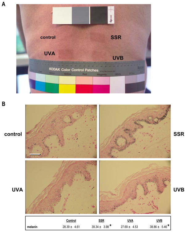
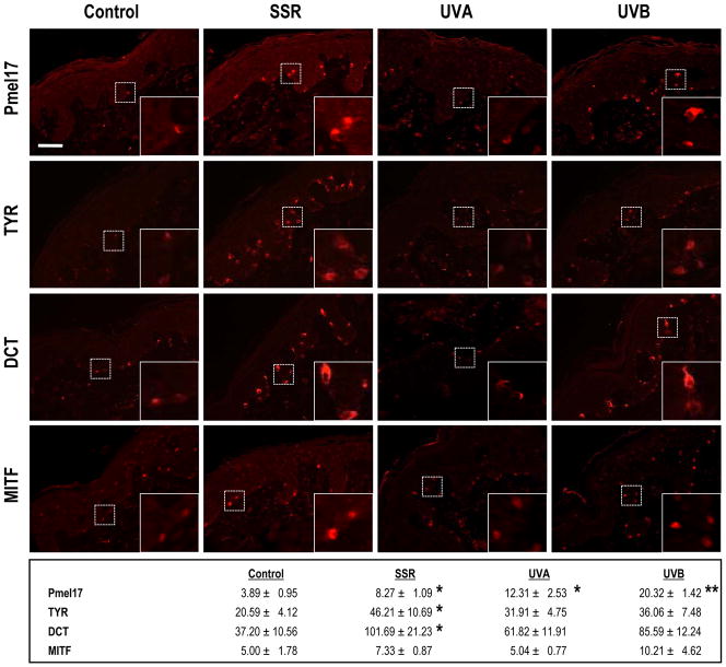
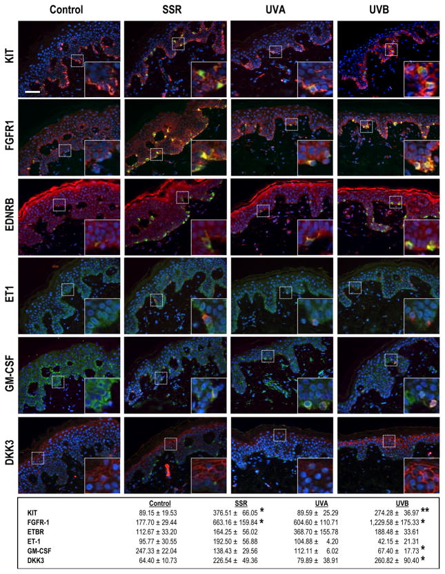
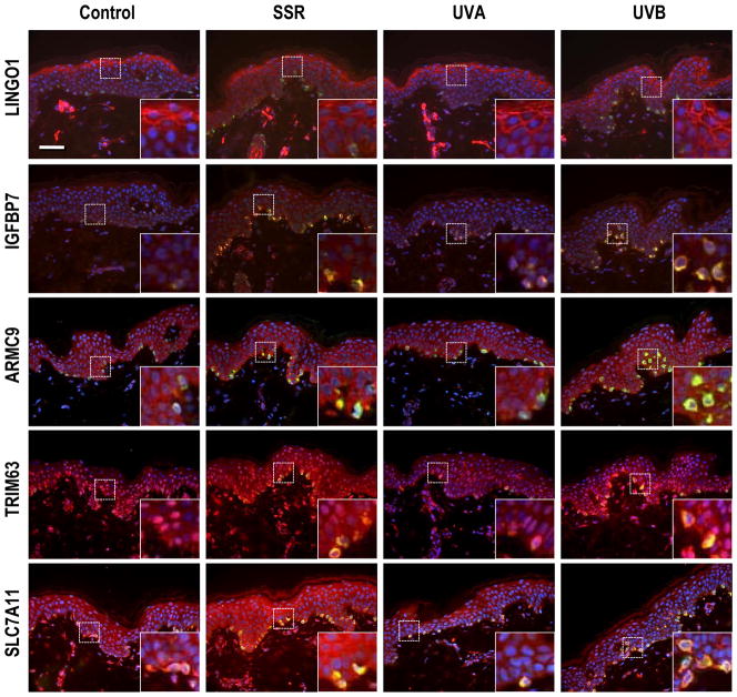
UV radiation is a major environmental factor that affects pigmentation in human skin and can eventually result in various types of UV-induced skin cancers. The effects of various wavelengths of UV on melanocytes and other types of skin cells in culture have been studied, but little is known about gene expression patterns in situ following in situ exposure of human skin to different types of UV (UVA and/or UVB). Paracrine factors expressed by keratinocytes and/or fibroblasts that affect skin pigmentation might be regulated differently by UV, as might their corresponding receptors expressed on melanocytes. To test the hypothesis that different mechanisms are involved in the pigmentary responses of the skin to different types of UV, we used immunohistochemical and whole human genome microarray analyses to characterize human skin in situ to examine how melanocyte-specific proteins and paracrine melanogenic factors are regulated by repetitive exposure to different types of UV compared with unexposed skin as a control. The results show that gene expression patterns induced by UVA or UVB are distinct-UVB eliciting dramatic increases in a large number of genes involved in pigmentation as well as in other cellular functions, whereas UVA had little or no effect on these. The expression patterns characterize the distinct responses of the skin to UVA or UVB, and identify several potential previously unidentified factors involved in UV-induced responses of human skin.
Conflict of interest statement
The authors state no conflict of interest.
Figures
References
-
- Alaluf S, Atkins D, Barrett K, Blount M, Carter N, Heath A. Ethnic variation in melanin content and composition in photoexposed and photoprotected human skin. Pigment Cell Res. 2002a;15:112–118. - PubMed
-
- Alaluf S, Atkins D, Barrett K, Blount M, Carter N, Heath A. The impact of epidermal melanin on objective measurements of human skin colour. Pigment Cell Res. 2002b;15:119–126. - PubMed
-
- Bancroft JD, Stevens A. Theory and Practice of Histological Techniques. Churchill Livingstone; New York: 1982.
-
- Bech-Thomsen N, Ravnborg L, Wulf HC. A quantitative study of the melanogenic effect of multiple suberythemal doses of different ultraviolet radiation sources. Photodermatol Photoimmunol Photomed. 1994;10:53–56. - PubMed
-
- Black HS, de Gruijl FR, Forbes PD, Cleaver JE, Ananthaswamy HN, De Fabo EC, Ullrich SE, Tyrrell RM. Photocarcinogenesis: an overview. Photochem Photobiol. 1997;40:29–47. - PubMed
Publication types
MeSH terms
Substances
Grants and funding
LinkOut - more resources
Full Text Sources
Other Literature Sources
Molecular Biology Databases
