

Automatic measurement of epithelium differentiation and classification of cervical intraneoplasia by computerized image analysis
- PMID: 20148100
- PMCID: PMC2819044
- DOI: 10.1186/1746-1596-5-7
Automatic measurement of epithelium differentiation and classification of cervical intraneoplasia by computerized image analysis
Abstract
Background: The feasibility of evaluating an objective grading of cervical intraneoplasia lesions (CIN) is attempted using an automatic computerized system able to measure several valuable parameters with special reference to epithelium differentiation.
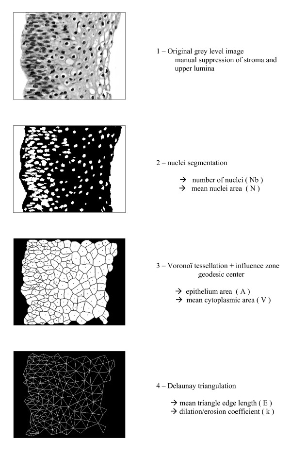
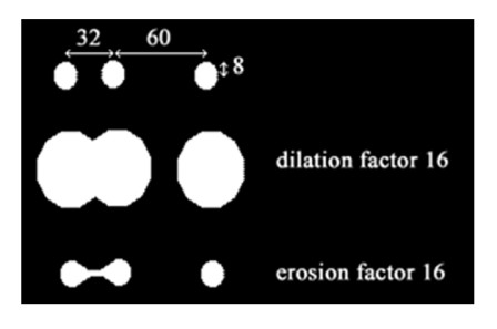
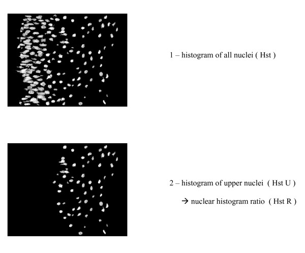
Methods: 4 groups of 10 images each were selected at random from 68 consensus images coming from 80 archival cervical biopsies, normal (n = 10), CIN 1 (n = 10), CIN 2 (n = 10), CIN 3 (n = 10). Representative images of lesions were captured from the microscopic slides and were analyzed using mathematical morphology, with special reference toVoronoï tessellation and Delaunay triangulation. Epithelium surface, nuclear and cytoplasm area, triangle edge and area, total and upper nuclear index were precisely measured in each lesion, and discriminant coefficients were calculated therewith. A dilation/erosion coefficient was automatically defined using triangle edge length and nuclear radius in order to measure the epithelium ratio of differentiation. A histogram ratio was also automatically established between total nuclei and upper nuclei on top of differentiated epithelium. With the latter two ratios added to the nucleo-cytoplasmic ratio, a cervical score able to classify CIN is proposed.
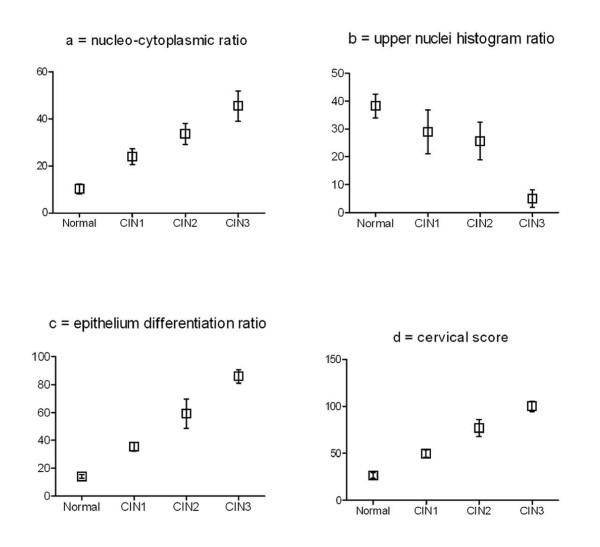
Results: There is a quasi-linear increase of mean cervical score values between normal epithelium and CIN 3: (27) for normal epithelium, (51) for CIN 1, (78) for CIN 2 and (100) for CIN 3, with significant differences (P < 0.05).
Conclusion: Our results highlight the possibility of applying a cervical score for the automatic grading of CIN lesions and thereby assisting the pathologist for improvement of grading. The automatic measure of epithelium differentiation ratio appears to be a new interesting parameter in computerized image analysis of cervical lesions.
Figures

Similar articles
-
An automated machine vision system for the histological grading of cervical intraepithelial neoplasia (CIN).J Pathol. 2000 Nov;192(3):351-62. doi: 10.1002/1096-9896(2000)9999:9999<::AID-PATH708>3.0.CO;2-I. J Pathol. 2000. PMID: 11054719
-
False negative colposcopy is associated with thinner cervical intraepithelial neoplasia 2 and 3.Gynecol Oncol. 2008 Jul;110(1):32-6. doi: 10.1016/j.ygyno.2008.03.003. Epub 2008 May 16. Gynecol Oncol. 2008. PMID: 18485462
-
Quantitative histopathological analysis of cervical intra-epithelial neoplasia sections: methodological issues.Cell Oncol. 2004;26(1-2):31-43. doi: 10.1155/2004/238769. Cell Oncol. 2004. PMID: 15371655 Free PMC article.
-
Coordinate expression of cytokeratin 8 and cytokeratin 17 immunohistochemical staining in cervical intraepithelial neoplasia and cervical squamous cell carcinoma: an immunohistochemical analysis and review of the literature.Gynecol Oncol. 2008 Mar;108(3):598-602. doi: 10.1016/j.ygyno.2007.11.042. Epub 2008 Jan 14. Gynecol Oncol. 2008. PMID: 18191996 Review.
-
[Precancerous lesions of the uterine cervix: morphology and molecular pathology].Pathologe. 2011 Nov;32 Suppl 2:242-54. doi: 10.1007/s00292-011-1517-0. Pathologe. 2011. PMID: 21909794 Review. German.
Cited by
-
Open source tools for management and archiving of digital microscopy data to allow integration with patient pathology and treatment information.Diagn Pathol. 2013 Feb 12;8:22. doi: 10.1186/1746-1596-8-22. Diagn Pathol. 2013. PMID: 23402499 Free PMC article.
-
Molecular characteristics and chromatin texture features in acute promyelocytic leukemia.Diagn Pathol. 2012 Jun 28;7:75. doi: 10.1186/1746-1596-7-75. Diagn Pathol. 2012. PMID: 22742960 Free PMC article.
-
Exploring automatic prostate histopathology image Gleason grading via local structure modeling.Annu Int Conf IEEE Eng Med Biol Soc. 2015;2015:2649-52. doi: 10.1109/EMBC.2015.7318936. Annu Int Conf IEEE Eng Med Biol Soc. 2015. PMID: 26736836 Free PMC article.
-
An intelligent clinical decision support system for patient-specific predictions to improve cervical intraepithelial neoplasia detection.Biomed Res Int. 2014;2014:341483. doi: 10.1155/2014/341483. Epub 2014 Apr 9. Biomed Res Int. 2014. PMID: 24812614 Free PMC article.
-
Fractal dimension of chromatin is an independent prognostic factor for survival in melanoma.BMC Cancer. 2010 Jun 5;10:260. doi: 10.1186/1471-2407-10-260. BMC Cancer. 2010. PMID: 20525386 Free PMC article.
References
-
- Mccluggage WG, Walsh MY, Thornton CM, Hamilton PW, Date A, Caughley LM, Bharucha H. Inter- and intra-observer variation in the histopathological reporting of cervical squamous intraepithelial lesions using a modified Bethesda grading system. Br J Obstet Gynaecol. 1998. pp. 206–210. - PubMed
-
- Malpica A, Matisic JP, Niekirk DV, Crum CP, Staerkel GA, Yamal JM, Guillaud MH, Cox DD, Atkinson EN, Adler-Storthz K, Poulin NM, Macaulay CA, Follen M. Kappa statistics to measure interrater and intraraterment for 1790 cervical biopsy specimens among twelve pathologists: qualitative histopathologic analysis and methodologic issues. Gynecol Oncol. 2005;3(Suppl 1):S38–52. doi: 10.1016/j.ygyno.2005.07.040. - DOI - PubMed
