

A patient with unilateral tibial aplasia and accessory scrotum: a pure coincidence or nonfortuitous association?
- PMID: 20148169
- PMCID: PMC2817541
- DOI: 10.1155/2010/898636
A patient with unilateral tibial aplasia and accessory scrotum: a pure coincidence or nonfortuitous association?
Abstract
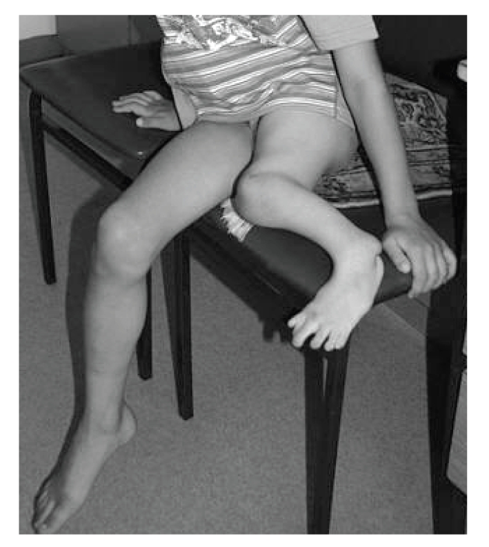
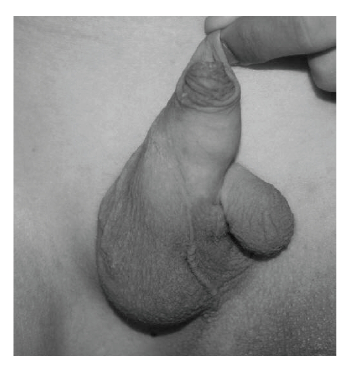
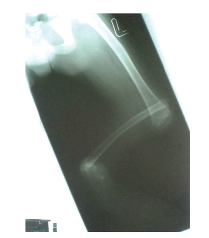
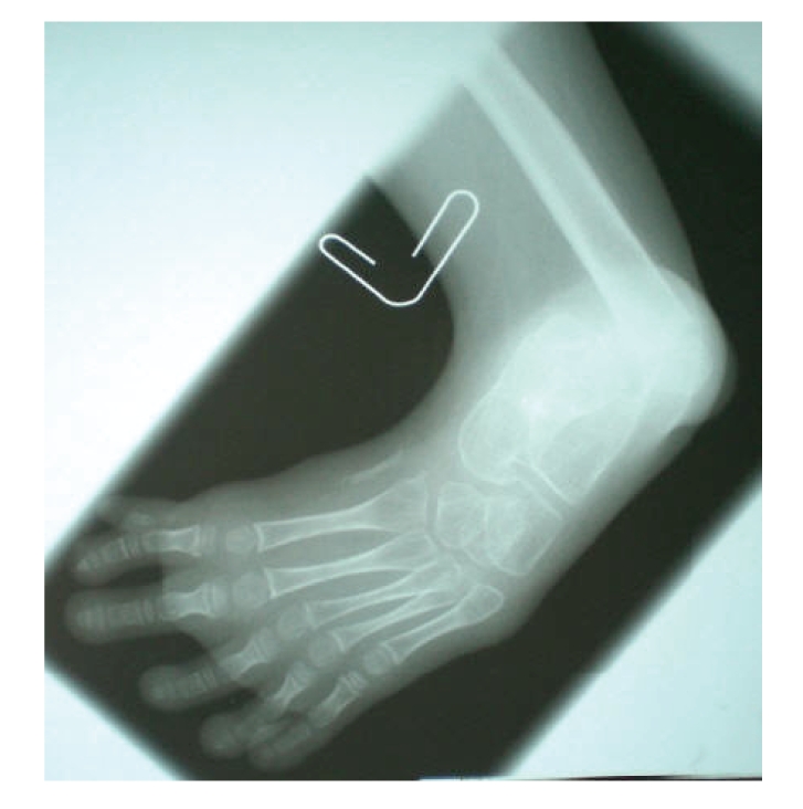
Tibial aplasia is an uncommon lower limb malformation that can occur isolated or be part of a more complex malformation pattern. We describe a 9-year-old boy born after uneventful pregnancy and delivery. Family history was negative for maternal diabetes and other malformations. The patient presented with left tibial aplasia and homolateral prexial foot polydactyly. He also displayed enamel dysplasia and bifid scotum with cryptorchidism. Literature review failed to identify a significant syndromic association between lower limb defects of the tibial type and the genital anomalies reported here. The combination of tibial aplasia with midline genital malformations further supports the hypothesis that the tibial ray development mirrors the morphogenetic process of the radial structures. Accordingly, the malformation pattern observed in the present patient may be pathogenetically explained by an insult occurring during late blastogenesis.
Figures
References
-
- Castori M, Rinaldi R, Cappellacci S, Grammatico P. Tibial developmental field defect is the most common lower limb malformation pattern in VACTERL association. American Journal of Medical Genetics A. 2008;146(10):1259–1266. - PubMed
-
- Jones D, Barnes J, Lloyd-Roberts GC. Congenital aplasia and dysplasia of the tibia with intact fibula: classification and management. Journal of Bone and Joint Surgery. British. 1978;60(1):31–39. - PubMed
-
- Slavotinek A, Clayton-Smith J, Kerr B. Unilateral tibial aplasia, pre-axial polysyndactyly, vertebral anomalies and imperforate anus. Clinical Dysmorphology. 1999;8(3):223–225. - PubMed
-
- Tüysüz B, Beker BD, Centel T, Üngür S, Lter O. Unilateral tibial agenesia with preaxial polysyndactyly and renal disorder in two patients: a new syndrome? Clinical Dysmorphology. 2001;10(1):37–40. - PubMed
-
- Evans JA, Greenberg CR. Tibial agenesis with radial ray and cardiovascular defects. Clinical Dysmorphology. 2002;11(3):163–169. - PubMed
Publication types
LinkOut - more resources
Full Text Sources
