. 2009 Apr;1(4):402-11.
doi: 10.18632/aging.100042.

MicroRNAs miR-146a/b negatively modulate the senescence-associated inflammatory mediators IL-6 and IL-8

Affiliations

MicroRNAs miR-146a/b negatively modulate the senescence-associated inflammatory mediators IL-6 and IL-8

Dipa Bhaumik et al. Aging (Albany NY). 2009 Apr.

Abstract

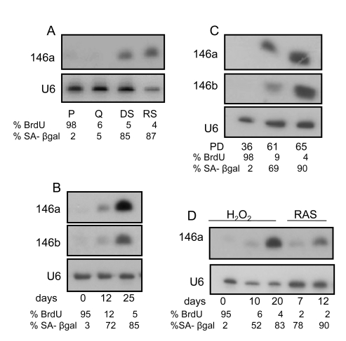
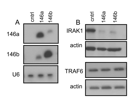
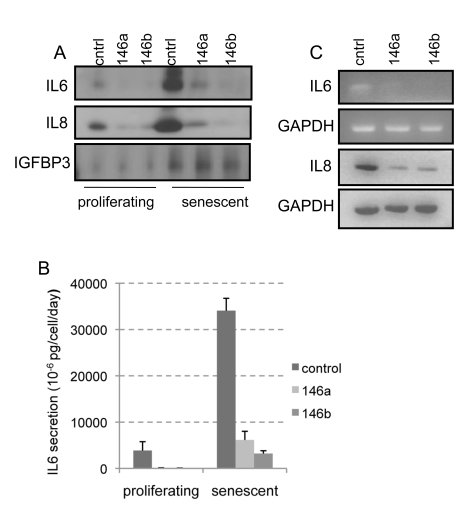
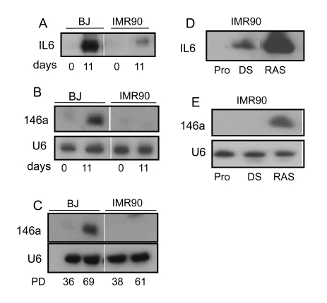
Senescence is a cellular program that irreversibly arrests the proliferation of damaged cells and induces the secretion of the inflammatory mediators IL- 6 and IL-8 which are part of a larger senescence associated secretory phenotype (SASP). We screened quiescent and senescent human fibroblasts for differentially expressed microRNAS (miRNAs) and found that miRNAs 146a and 146b (miR-146a/b) were significantly elevated during senescence. We suggest that delayed miR-146a/b induction might be a compensatory response to restrain inflammation. Indeed, ectopic expression of miR-146a/b in primary human fibroblasts suppressed IL-6 and IL-8 secretion and downregulated IRAK1, a crucial component of the IL-1 receptor signal transduction pathway. Cells undergoing senescence without induction of a robust SASP did not express miR-146a/b. Further, IL-1alpha neutralizing antibodies abolished both miR-146a/b expression and IL-6 secretion. Our findings expand the biological contexts in which miRNA-146a/b modulates inflammatory responses. They suggest that IL-1 receptor signaling initiates both miR-146a/b upregulation and cytokine secretion, and that miR-146a/b is expressed in response to rising inflammatory cytokine levels as part of a negative feedback loop that restrains excessive SASP activity.

Keywords: DNA damage; IL-1α; IL-6; IL-8; inflammation; miRNA.


Conflict of interest statement

There is no conflict of interest for any of the authors.

Figures

Figure 1.
Figure 1.. miR146a/b expression increases in senescent HCA2 fibroblasts.
(A) Northern blot analysis of total RNA prepared from proliferating (P), quiescent (Q), damage (bleomycin)-induced senescent (DS) and replicatively senescent (RS) HCA2 cells. We analyzed 10 μg of RNA from P, Q and DS cells, but 5 μg of RNA from RS cells. After separation and transfer to membranes, the blots were probed for miR-146a. Equal RNA loading was confirmed by probing for the small RNA species U6. Values for the percentage of cells incorporating bromodeoxyuridine (% BrdU) or expressing the sensecence-associated beta-galactosidase (% SA-β-gal) are indicated below each lane. (B) Northern blot analysis of RNA from DS cells. Cells were harvested for RNA at the days indicated after cells were induced to senesce by bleomycin. The blot was initially probed for miR-146a, then stripped and reprobed for miR-146b. The proliferation levels (% BrdU) and % cells that express the SA-β-gal are indicated. (C) Northern blot analysis of replicatively senescencing cells. Cells were harvested at the PD (population doubling level) indicated below the figure. The proliferation levels (% BrdU) and % cells that express the SA-β-gal are indicated. (D) Northern blot analysis of cells treated with H2O2 (0.1 mM for 2 h) or infected with the lentivirus expressing oncogenic RASV12. Cells were harvested for RNA at the indicated days after treatment. The proliferation levels (% BrdU) and % cells that express the SA-β-gal are indicated.
Figure 2.
Figure 2.. IRAK1 but not TRAF6 levels are reduced in HCA2 cells overexpressing miR146a and miR 146b.
(A) Northern blot analysis of total RNA prepared from control (insertless virus-infected) HCA2 cells (cntrl), cells infected with a miR-146a-expressing virus (146a) and cells infected with a miR-146b-expressing virus (146b). 8 μg of total RNA was loaded in each lane. (B) Western blot analysis of total protein lysates prepared from proliferating cells (cntrl, PD32), or cells overexpressing miR-146a or miR-146b, and analyzed for IRAK1 (top panel) and TRAF6 (bottom panel). Actin protein levels served as a loading control.
Figure 3.
Figure 3.. Overexpression of miR-146a/b suppresses basal and senescence-associated secretion of IL-6 and IL-8 in HCA2 cells.
(A) Western blot analyses of TCA-precipitated proteins prepared from CM collected over 24 h from cells infected with the lentivirus backbone (cntrl) or lentiviruses expressing miR-146a (146a) or miR-146b (146b). The blot was analyzed for IL-6, IL-8 and IGFBP3. Equal loading was based on cell number prior to collection of CM and IGFBP3 levels. Proliferating indicates cells described in Figure 2A. The same cells were treated with bleomycin and CM was harvested 11 days later (senescent). (B) IL-6 in CM from the cell populations described in Fig 3A was measured by ELISA. The data are reported as 10-6 pg per cell per day. (C) RT-PCR analysis of transcript levels of IL-6 and IL-8 in miR-146a/b-overexpressing cells. RNA collected from proliferating cells was used as the control (cntrl).
Figure 4.
Figure 4.. miR-146a/b increase in senescent fibroblasts that secrete high levels of inflammatory cytokines.
(A) & (B) Proliferating BJ (PD 36) and IMR90 (PD 38) cells were treated with bleomycin to induce senescence. CM and RNA were harvested 11 d later. (A) Western analysis for secreted IL-6. (B) Northern analysis for miR-146a. (C) Northern analysis for miR-146a levels in replicatively senescent BJ and IMR90 cells. The PD levels at which cells were harvested for analysis is given below each lane. BJ cells reach complete senescence after approximately 70 PDs, whereas IMR90 cells are nearly completely senescent by PD61. (D) & (E) Proliferating IMR90 cells (PD40) were either untreated (Pro), treated with bleomycin (DS) or infected with the lentivirus expressing oncogenic RAS (RAS). CM and RNA were collected 11 days after treatment or infection. (D) Western analysis for IL-6 in CM. (E) Northern analysis for miR-146a and U6 (control) levels.
Figure 5.
Figure 5.. IL-1α upregulates miR-146a in senescent cells.
(A) Northern analysis for miR-146a levels in damage-induced senescent HCA2 cells treated with neutralizing antibodies against IL1-α and IL1-β. HCA2 cells (PD 35) were used and induced to senesce by treatment with bleomycin. Cells were harvested for RNA 11 days later. Details of the procedure are described in ‘Experimental Procedures. (B) Western analysis for IL-6 in damage-induced senescent HCA2 cells treated with neutralizing antibodies to IL-1α and IL-1β. CM were harvested 11 days after bleomycin treatment. (C) Model for the role of miR-146a/b in senescent cells: In response to a high SASP (right branch), IL-1α interacts with the IL-1α receptor (IL-1αR) and the signaling pathway that involves IRAK1 is fully activated. This activation leads to the well-documented activation of the transcription factor NFкB and production of IL-6, IL-8 and also miRNA-146a/b. miRNA-146a/b is a component of a negative feedback loop and acts to downregulate the levels of IRAK1, hence restraining the levels of IL-6 and IL8. However, in response to a low SASP (left branch), the signaling pathway is not sufficiently activated. Thus there is a low level of IL-6 and IL-8 secretion and miRNA-146a/b is not upregulated.

Similar articles

Cited by

References

    1. Campisi J. Senescent cells, tumor suppression, and organismal aging: good citizens, bad neighbors. Cell. 2005;120:513–522. - PubMed
    1. Campisi J, d'Adda di Fagagna F. Cellular senescence: when bad things happen to good cells. Nat Rev Mol Cell Biol. 2007;8:729–740. - PubMed
    1. Lundberg AS, Hahn WC, Gupta P, Weinberg RA. Genes involved in senescence and immortalization. Curr Opin Cell Biol. 2000;12:705–709. - PubMed
    1. Serrano M, Blasco MA. Putting the stress on senescence. Curr Opin Cell Biol. 2001;13:748–753. - PubMed
    1. Demidenko ZN, Blagosklonny MV. Growth stimulation leads to cellular senescence when the cell cycle is blocked. Cell Cycle. 2008;7:3355–3361. - PubMed

Publication types

MeSH terms

LinkOut - more resources