Review
. 2010 Jun;67(12):1957-70.
doi: 10.1007/s00018-010-0279-9. Epub 2010 Feb 11.

Polo-box domain: a versatile mediator of polo-like kinase function

Affiliations
Review

Polo-box domain: a versatile mediator of polo-like kinase function

Jung-Eun Park et al. Cell Mol Life Sci. 2010 Jun.

Abstract

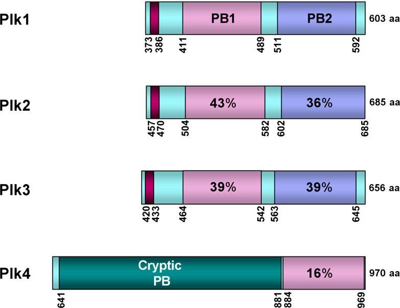
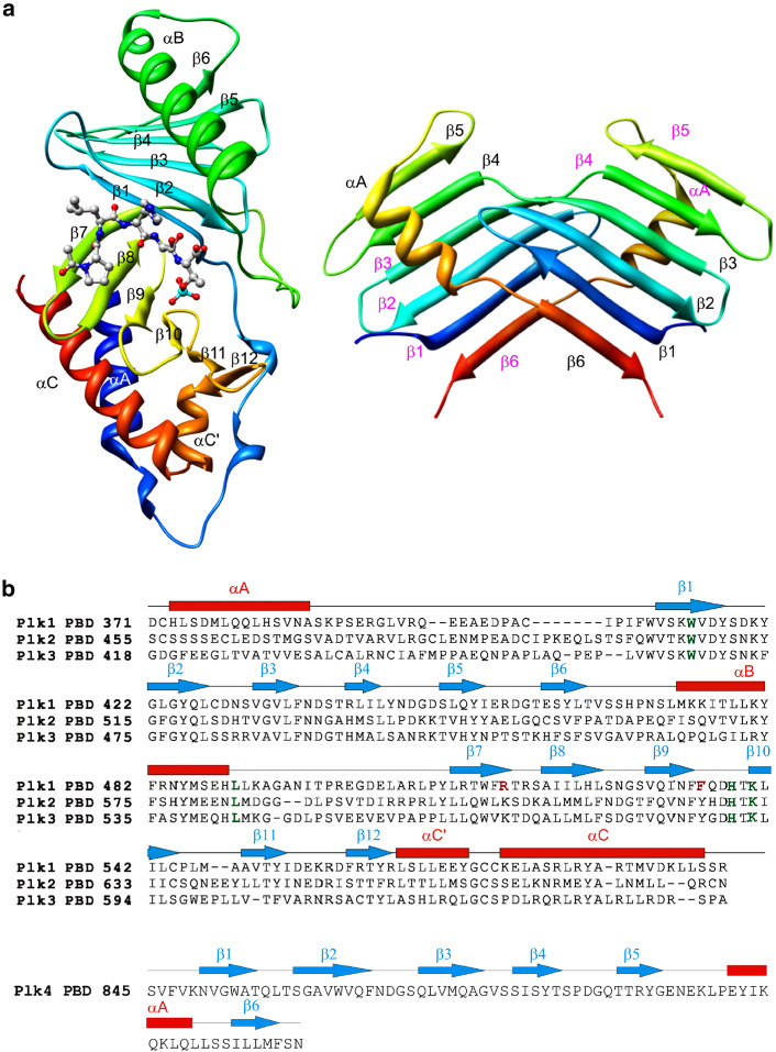
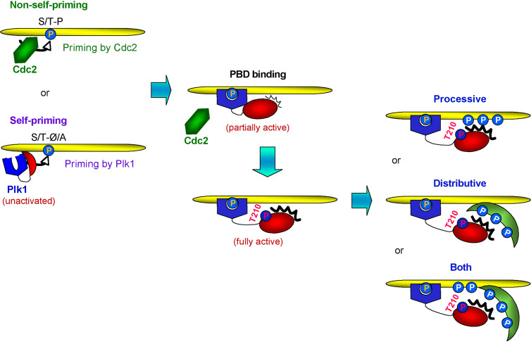
Members of the polo subfamily of protein kinases have emerged as important regulators in diverse aspects of the cell cycle and cell proliferation. A large body of evidence suggests that a highly conserved polo-box domain (PBD) present in the C-terminal non-catalytic region of polo kinases plays a pivotal role in the function of these enzymes. Recent advances in our comprehension of the mechanisms underlying mammalian polo-like kinase 1 (Plk1)-dependent protein-protein interactions revealed that the PBD serves as an essential molecular mediator that brings the kinase domain of Plk1 into proximity with its substrates, mainly through phospho-dependent interactions with its target proteins. In this review, current understanding of the structure and functions of PBD, mode of PBD-dependent interactions and substrate phosphorylation, and other phospho-independent functions of PBD are discussed.


Figures

Fig. 1
Fig. 1
Subcellular localization of Plk1 during the cell cycle. Plk1 localizes to the centrosomes as early as S phase. As Plk1 becomes more abundant during the late stages of the cell cycle, Plk1 localization to the centrosomes (arrows) and kinetochores (dotted red fluorescent signals) are manifest. In anaphase, a fraction of Plk1 relocalizes to the spindle midzone, which then condenses to a midbody in telophase
Fig. 2
Fig. 2
Schematic diagram of the PBDs from human Plk1–4. Sequence identities among PB1s (light pink) and PB2s (light blue) are given in percentages. A short sequence (burgundy) upstream of PB1 represent a short alpha-helix polo-cap denoted as αA in Fig. 3. The total lengths of Plk1–4 are indicated in amino acid residue numbers on the right. The numbers indicate the positions of the amino acid residues in a given gene. Cryptic PB indicates a region of Plk4 that is also sufficient for dimerization [2, 62]
Fig. 3
Fig. 3
Structures of the PBDs from human Plk1–4. a Comparison of the crystal structures of PBD1 and PBD4. The structure of PBD1 is shown in complex with PLHSpT drawn as a ball and stick representation [61]. The structure of PBD4 is adopted from Leung et al. [62]. Notations for α-helices and β-strands are given in order. b The sequence alignment of the PBDs from Plk1–3. Notations for α-helices and β-strands are done in the same manner as in (a). Four conserved residues (W414, L490, H538, and K540) from the PBD1 important for binding to the MQSpTPL-containing optimal peptide are highlighted in green. Two unique residues (R516 and F535) critical for accommodating the N-terminal Pro of PLHSpT are highlighted in red. PBD4 does not exhibit a significant level of homology with PBD1–3, and thus is shown separately
Fig. 4
Fig. 4
Schematic diagram for Plk1 activation, PBD1-dependent binding, and substrate phosphorylation. Generation of PBD1-binding targets is achieved either by a Pro-directed kinase such as Cdc2 (nonself-priming) or by Plk1 itself (self-priming). Upon binding to a phosphorylated target through PBD1, Plk1 becomes partially active via physical dissociation of its kinase domain from the PBD1. Phosphorylation of Plk1 at T210 by an upstream kinase such as Aurora A/Bora [80, 86] fully activates the enzyme prior to G2/M transition. Once Plk1 binds to a phosphorylated target, Plk1 phosphorylates either the same protein binding to its PBD1 (processive phosphorylation) or a third protein associating with the PBD1-binding target (distributive phosphorylation). These two phosphorylation models are not mutually exclusive and may operate in a concerted manner (both)
Fig. 5
Fig. 5
Phospho-dependent interactions between PBD1 and its binding targets are critical but not essential for the subcellular localization of Plk1. HeLa cells infected with adenoviruses expressing either full-length EGFP-Plk1 (WT) or EGFP-Plk1 (H538A K540M) (AM) were fixed and imaged by confocal microscopy (a). The fluorescent intensities of Plk1 signals at the centrosomes, kinetochores, midzone, and midbody were analyzed (b)

References

    1. Sunkel CL, Glover DM. polo, a mitotic mutant of Drosophila displaying abnormal spindle poles. J Cell Sci. 1988;89:25–38. - PubMed
    1. Archambault V, Glover DM. Polo-like kinases: conservation and divergence in their functions and regulation. Nat Rev Mol Cell Biol. 2009;10:265–275. doi: 10.1038/nrm2653. - DOI - PubMed
    1. Barr FA, Sillje HH, Nigg EA. Polo-like kinases and the orchestration of cell division. Nat Rev Mol Cell Biol. 2004;5:429–440. doi: 10.1038/nrm1401. - DOI - PubMed
    1. Winkles JA, Alberts GF. Differential regulation of polo-like kinase 1, 2, 3, and 4 gene expression in mammalian cells and tissues. Oncogene. 2005;24:260–266. doi: 10.1038/sj.onc.1208219. - DOI - PubMed
    1. Clay FJ, McEwen SJ, Bertoncello I, Wilks AF, Dunn AR. Identification and cloning of a protein kinase-encoding mouse gene, Plk, related to the polo gene of Drosophila . Proc Natl Acad Sci USA. 1993;90:4882–4886. doi: 10.1073/pnas.90.11.4882. - DOI - PMC - PubMed

MeSH terms

LinkOut - more resources