

Astrogliosis in CNS pathologies: is there a role for microglia?
- PMID: 20148316
- PMCID: PMC3629545
- DOI: 10.1007/s12035-010-8098-4
Astrogliosis in CNS pathologies: is there a role for microglia?
Abstract
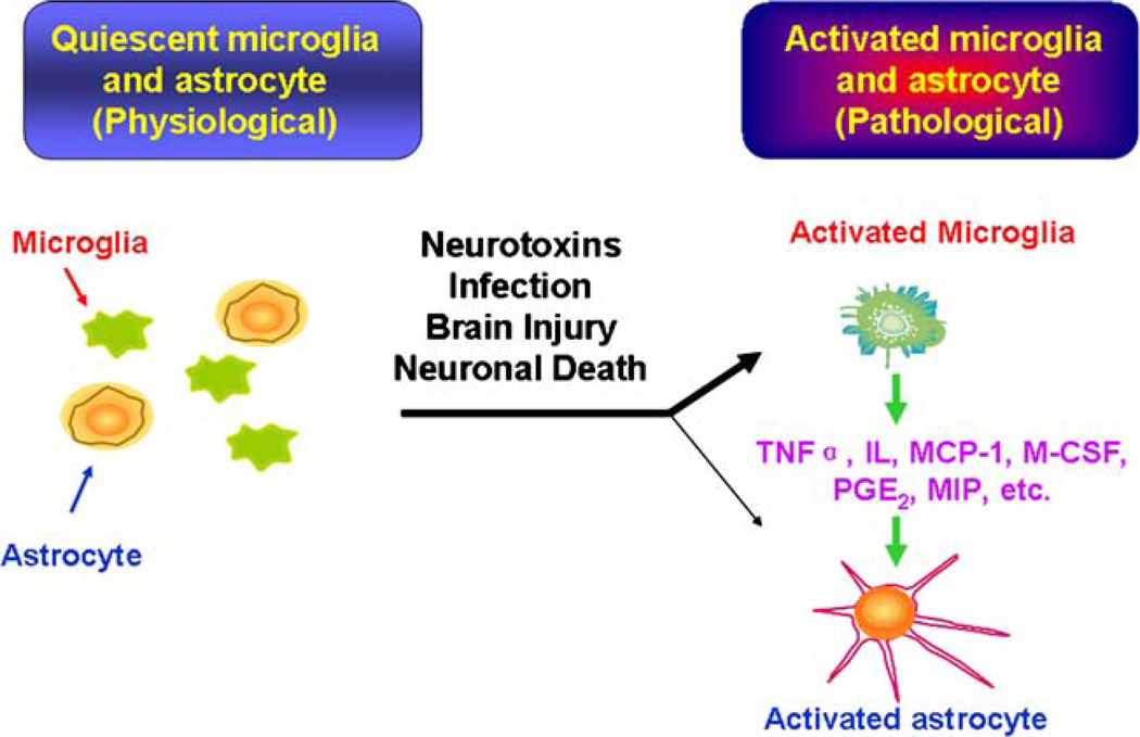
Astrogliosis, a cellular reaction with specific structural and functional characteristics, represents a remarkably homotypic response of astrocytes to all kinds of central nervous system (CNS) pathologies. Astrocytes play diverse functions in the brain, both harmful and beneficial. Mounting evidence indicates that astrogliosis is an underlying component of a diverse range of diseases and associated neuropathologies. The mechanisms that lead to astrogliosis are not fully understood, nevertheless, damaged neurons have long been reported to induce astrogliosis and astrogliosis has been used as an index for underlying neuronal damage. As the predominant source of proinflammatory factors in the CNS, microglia are readily activated under certain pathological conditions. An increasing body of evidence suggests that release of cytokines and other soluble products by activated microglia can significantly influence the subsequent development of astrogliosis and scar formation in CNS. It is well known that damaged neurons activate microglia very quickly, therefore, it is possible that activated microglia contribute factors/mediators through which damaged neuron induce astrogliosis. The hypothesis that activated microglia initiate and maintain astrogliosis suggests that suppression of microglial overactivation might effectively attenuate reactive astrogliosis. Development of targeted anti-microglial activation therapies might slow or halt the progression of astrogliosis and, therefore, help achieve a more beneficial environment in various CNS pathologies.
Figures
Similar articles
-
Reciprocal modulation between microglia and astrocyte in reactive gliosis following the CNS injury.Mol Neurobiol. 2013 Dec;48(3):690-701. doi: 10.1007/s12035-013-8460-4. Epub 2013 Apr 24. Mol Neurobiol. 2013. PMID: 23613214 Free PMC article. Review.
-
Astrogliosis in the adult and developing CNS: is there a role for proinflammatory cytokines?Neurotoxicology. 2001 Oct;22(5):607-18. doi: 10.1016/s0161-813x(01)00032-8. Neurotoxicology. 2001. PMID: 11770882 Review.
-
Distribution of the immune inhibitory molecules CD200 and CD200R in the normal central nervous system and multiple sclerosis lesions suggests neuron-glia and glia-glia interactions.J Neuropathol Exp Neurol. 2009 Feb;68(2):159-67. doi: 10.1097/NEN.0b013e3181964113. J Neuropathol Exp Neurol. 2009. PMID: 19151626
-
CNTF regulation of astrogliosis and the activation of microglia in the developing rat central nervous system.Brain Res. 1995 Jul 10;685(1-2):55-67. doi: 10.1016/0006-8993(95)00411-i. Brain Res. 1995. PMID: 7583254
-
Astrocytes: biology and pathology.Acta Neuropathol. 2010 Jan;119(1):7-35. doi: 10.1007/s00401-009-0619-8. Epub 2009 Dec 10. Acta Neuropathol. 2010. PMID: 20012068 Free PMC article. Review.
Cited by
-
Induction of a Proinflammatory Response in Cortical Astrocytes by the Major Metabolites Accumulating in HMG-CoA Lyase Deficiency: the Role of ERK Signaling Pathway in Cytokine Release.Mol Neurobiol. 2016 Aug;53(6):3586-3595. doi: 10.1007/s12035-015-9289-9. Epub 2015 Jun 23. Mol Neurobiol. 2016. PMID: 26099308
-
Inflammatory Foreign Body Response Induced by Neuro-Implants in Rat Cortices Depleted of Resident Microglia by a CSF1R Inhibitor and Its Implications.Front Neurosci. 2021 Mar 26;15:646914. doi: 10.3389/fnins.2021.646914. eCollection 2021. Front Neurosci. 2021. PMID: 33841088 Free PMC article.
-
Impaired Learning and Memory Ability Induced by a Bilaterally Hippocampal Injection of Streptozotocin in Mice: Involved With the Adaptive Changes of Synaptic Plasticity.Front Aging Neurosci. 2021 Mar 1;13:633495. doi: 10.3389/fnagi.2021.633495. eCollection 2021. Front Aging Neurosci. 2021. PMID: 33732137 Free PMC article.
-
Liposomal clodronate selectively eliminates microglia from primary astrocyte cultures.J Neuroinflammation. 2012 May 31;9:116. doi: 10.1186/1742-2094-9-116. J Neuroinflammation. 2012. PMID: 22651847 Free PMC article.
-
Recombinant adeno-associated viral (rAAV) vectors mediate efficient gene transduction in cultured neonatal and adult microglia.J Neurochem. 2016 Jan;136 Suppl 1(0 1):49-62. doi: 10.1111/jnc.13081. Epub 2015 Mar 20. J Neurochem. 2016. PMID: 25708596 Free PMC article.
References
-
- Bogen IL, Risa O, Haug KH, Sonnewald U, Fonnum F, Walaas SI. Distinct changes in neuronal and astrocytic amino acid neurotransmitter metabolism in mice with reduced numbers of synaptic vesicles. J Neurochem. 2008;105:2524–2534. - PubMed
-
- Araque A. Astrocytes process synaptic information. Neuron Glia Biol. 2008;4:3–10. - PubMed
-
- Hu R, Cai WQ, Wu XG, Yang Z. Astrocyte-derived estrogen enhances synapse formation and synaptic transmission between cultured neonatal rat cortical neurons. Neuroscience. 2007;144:1229–1240. - PubMed
Publication types
MeSH terms
Substances
Grants and funding
LinkOut - more resources
Full Text Sources
Other Literature Sources
Research Materials
