. 2010 Feb;26(1):1-10.
doi: 10.1089/jop.2009.0025.

Lovastatin inhibits the thrombin-induced loss of barrier integrity in bovine corneal endothelium

Affiliations

Lovastatin inhibits the thrombin-induced loss of barrier integrity in bovine corneal endothelium

Mahesh Shivanna et al. J Ocul Pharmacol Ther. 2010 Feb.

Abstract

Purpose: Increased actomyosin contraction of the dense band of actin cytoskeleton at the apical junctional complex (perijunctional actomyosin ring, PAMR) breaks down the barrier integrity of corneal endothelium. This study has investigated the efficacy of statins, which inhibit activation of RhoA, in opposing the thrombin-induced loss of barrier integrity of monolayers of cultured bovine corneal endothelium.

Methods: Myosin light chain (MLC) phosphorylation, a biochemical measure of actomyosin contraction, was assayed by urea-glycerol gel electrophoresis, followed by western blot analysis. The locus of MLC phosphorylation and changes in the organization of the PAMR were visualized by immunostaining. Phosphorylation of MYPT1, a regulatory subunit of myosin light-chain phosphatase (MLCP), was assessed by Western blot analysis to determine down-regulation of RhoA. The barrier integrity was assessed in terms of trans-endothelial electrical resistance (TER), and further confirmed by determining permeability to FITC dextran (10 kDa) and distribution of ZO-1, a marker of tight junctional assembly.

Results: Lovastatin, a prototype of lipophilic statins, induced MLC dephosphorylation under basal conditions. It opposed increase in phosphorylation of MLC and MYPT1 in response to thrombin and nocodazole, agents known to activate RhoA in the endothelium. Pretreatment with the statin opposed the thrombin- and nocodazole-induced disruption of the PAMR and the thrombin-induced decline in TER. Lovastatin also opposed the thrombin- and nocodazole-induced increase in permeability to FITC dextran and redistribution of ZO-1. However, upon supplementation with GGPP (geranylgeranyl pyrophosphate), lovastatin failed to oppose the effects of thrombin and nocodazole on the PAMR, ppMLC, and ZO-1 distribution.

Conclusions: Lovastatin attenuates RhoA activation in the corneal endothelium presumably by reducing its isoprenylation. This underlies the suppression of the thrombin-induced loss in barrier integrity of the corneal endothelium.


Figures

<b>FIG. 1.</b>
FIG. 1.
Effect of lovastatin on myosin light chain (MLC) phosphorylation. MLC phosphorylation was assayed by urea–glycerol gel electrophoresis, followed by western blotting. Typical blot depicting changes in status of MLC phosphorylation (A) (NP, non-phosphorylated MLC; P, monophosphorylated MLC; PP, diphosphorylated MLC). Untreated cells (C) show a basal level of MLC phosphorylation. Treatment with 10 μM lovastatin (L) for 24 h induces MLC dephosphorylation. Exposure to 2 U/mL thrombin (T) for 2 min induces increased MLC phosphorylation. Pretreatment with lovastatin opposes MLC phosphorylation induced by thrombin. Bar graph of densitometric analysis (B) of the representative experiments (N = 3) shown in panel A expressed as %pMLC. Lovastatin significantly opposes thrombin-induced MLC phosphorylation. *Significantly greater than control (P < 0.05). **Significantly less than thrombin treatment (P < 0.05).
<b>FIG. 2.</b>
FIG. 2.
Influence of lovastatin on the localization of diphosphorylated myosin light chain (ppMLC). The localization of ppMLC was ascertained by immunostaining, followed by imaging with an epifluorescence microscope. The images are representative of 3 independent experiments. In untreated cells (A), ppMLC staining in the cell periphery is moderate, indicating a basal level of MLC phosphorylation. Treatment with 10 μM lovastatin for 24 h (B) reduces ppMLC when compared to untreated cells, indicating MLC dephosphorylation. Exposure to 2 U/mL of thrombin for 2 min (C) increases the punctate localization of ppMLC in the cell periphery, indicating increased MLC phosphorylation. Pretreatment with lovastatin (D) opposes the thrombin response. Upon treatment with 10 μM geranylgeranyl pyrophosphate (GGPP) for 24 h (E), the localization of ppMLC is similar to that of untreated cells. On co-treatment of 10 μM GGPP with 10 μM lovastatin for 24 h followed by exposure to thrombin (F), lovastatin fails to oppose thrombin-induced increase in ppMLC.
<b>FIG. 3.</b>
FIG. 3.
Effect of lovastatin on nocodazole-induced myosin light chain (MLC) phosphorylation. Western blot depicting changes in MLC phosphorylation in response to nocodazole with or without the presence of lovastatin (A). Exposure to 2 μM nocodazole (N) for 30 min induces increased MLC phosphorylation when compared to untreated cells (C). Pretreatment with lovastatin (L) opposes nocodazole-induced MLC phosphorylation. Bar graph of densitometric analysis (B) of the representative experiments (N = 3) shown in panel A. Lovastatin significantly opposes nocodazole-induced MLC phosphorylation. *Significantly greater than control (P < 0.05). **Significantly less than nocodazole treatment (P < 0.05).
<b>FIG. 4.</b>
FIG. 4.
Influence of lovastatin on nocodazole-induced changes in localization of diphosphorylated myosin light chain (ppMLC). The images are representative of 3 independent experiments. In untreated cells (A), the localization of ppMLC in the cortical region of the cell is less dense, indicative of a basal level of MLC phosphorylation. Treatment with 10 μM lovastatin for 24 h (B) reduces ppMLC, correlating with MLC dephosphorylation. Exposure to 2 μM nocodazole for 30 min (C) increases ppMLC in the cortical areas, which is suggestive of increased MLC phosphorylation. Pretreatment with lovastatin (D) opposes the nocodazole response. Upon treatment with 10 μM geranylgeranyl pyrophosphate (GGPP) for 24 h (E), the staining for ppMLC is similar to that of untreated cells. On co-treatment of 10 μM GGPP with 10 μM lovastatin for 24 h (F), lovastatin fails to oppose nocodazole-induced increase in ppMLC.
<b>FIG. 5.</b>
FIG. 5.
Effect of lovastatin on organization of cortical actin. The changes in organization of cortical actin were ascertained by staining for F-actin, followed by imaging with fluorescence microscopy. The images are representative of 3 independent experiments. In untreated cells (A), the characteristic organization of cortical actin in the form of perijunctional actomyosin ring (PAMR) is evident (shown by arrow). Upon treatment with 10 μM lovastatin alone for 24 h (B), the organization of actin cytoskeleton remains largely unaltered. Exposure to 2 U/mL thrombin for 5 min (C) induces disruption of the cortical actin and formation of inter-endothelial gaps (shown by arrows). Pretreatment with lovastatin (D) opposes thrombin-induced disruption of PAMR. Upon co-treatment of lovastatin with 10 μM geranylgeranyl pyrophosphate (GGPP) for 24 h (E), lovastatin fails to oppose thrombin-induced disruption of PAMR. Treatment with 2 μM nocodazole for 30 min (F) also induces disruption of PAMR (shown by arrows). Pretreatment with lovastatin opposes the nocodazole (G) response on PAMR. However, upon co-treatment of lovastatin with 10 μM GGPP for 24 h (H), lovastatin fails to oppose the influence of nocodazole on PAMR.
<b>FIG. 6.</b>
FIG. 6.
Influence of lovastatin on phosphorylation status of MYPT1, a regulatory subunit of myosin light-chain phosphatase (MLCP). Phosphorylation of MYPT1 at Thr853 was determined using a phospho-specific antibody. Treatment with 2 U/mL thrombin for 2 min or 2 μM nocodazole for 30 min increased phosphorylation of MYPT1 (Lanes 2 and 5, respectively). Upon treatment with 10 μM lovastatin for 24 h, the MYPT1 phosphorylation is suppressed when compared to untreated cells (Lane 3 vs. Lane 1). Pretreatment with lovastatin opposes the increase in MYPT1 phosphorylation induced by thrombin and nocodazole (Lanes 4 and 6, respectively).
<b>FIG. 7.</b>
FIG. 7.
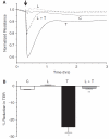
Effect of lovastatin on trans-endothelial electrical resistance (TER). The changes in TER were monitored by ECIS. In untreated cells (control), the TER remains relatively constant (A). Exposure to 3.5 U/mL thrombin induces an immediate decline in TER that fails to recover to baseline, even after nearly 3 h. Treatment with 10 μM lovastatin for 20 h slightly increases TER when compared to control. Pretreatment with 10 μM lovastatin for 20 h attenuates the extent of reduction in TER by thrombin and also facilitates faster recovery to baseline. Bar graph (B) of the peak response (<10 min) of representative experiments (N = 6) shown in Panel A. Lovastatin significantly opposes thrombin-induced reduction in TER. *Significantly greater than control (P < 0.001). **Significantly less than thrombin treatment (P < 0.001).
<b>FIG. 8.</b>
FIG. 8.
Influence of lovastatin on the localization of ZO-1, a marker of tight junctional assembly, in presence of thrombin. The localization of ZO-1 was ascertained by immunostaining, followed by imaging with fluorescence microscopy. The images are representative of 3 independent experiments. In untreated cells (A), ZO-1 localization is continuous and uniform throughout the cell periphery. Upon treatment with lovastatin alone (B), the localization of ZO-1 is unaltered. Treatment with 3.5 U/mL thrombin for 30 min (C) induces dispersion of ZO-1, indicating disorganization at the level of TJ (shown by arrows). Pretreatment with 10 μM lovastatin for 20 h (D) opposes thrombin response. Treatment with 10 μM geranylgeranyl pyrophosphate (GGPP) for 20 h (E) does not disturb the localization of ZO-1 and is similar to that of untreated cells. However, upon co-treatment of lovastatin with 10 μM GGPP for 24 h (F), lovastatin fails to oppose thrombin-induced dispersion of ZO-1.
<b>FIG. 9.</b>
FIG. 9.
Effect of lovastatin on permeability in presence of thrombin. The changes in permeability were ascertained by quantifying the flux of FITC dextran across cells grown on porous culture inserts. Treatment with 3.5 U/mL thrombin significantly increases permeability when compared to untreated cells. Pretreatment with 10 μM lovastatin for 20 h significantly attenuates thrombin response. The increase in permeability by lovastatin itself is insignificant compared to control. The data shown are representative of 8 independent experiments. *Significantly greater than control (P < 0.001). **Significantly less than thrombin treatment (P < 0.001).
<b>FIG. 10.</b>
FIG. 10.
Influence of lovastatin on ZO-1, a marker of tight junctional assembly, localization in presence of nocodazole. The images shown are representative of 3 independent experiments. In untreated cells (A), continuous peripheral pattern of ZO-1 localization is evident. Treatment with 10 μM lovastatin for 20 h (B) does not alter the pattern of ZO-1 localization and is similar to those of untreated cells. Treatment with 2 μM nocodazole for 30 min (C) induces discontinuities in ZO-1 localization (shown by arrows). Pretreatment with lovastatin (D) opposes nocodazole response. On treatment with 10 μM geranylgeranyl pyrophosphate (GGPP) for 20 h (E), the localization of ZO-1 is similar to that of untreated cells. However, upon co-treatment of lovastatin with 10 μM GGPP for 24 h (F), lovastatin fails to oppose nocodazole-induced dispersion of ZO-1.
<b>FIG. 11.</b>
FIG. 11.
Effect of lovastatin on permeability in presence of nocodazole. Treatment with 2 μM nocodazole significantly increases permeability when compared to control. Pretreatment with lovastatin significantly attenuates nocodazole response. The data shown are representative of 8 independent experiments. *Significantly greater than control (P < 0.001). **Significantly less than nocodazole treatment (P < 0.001).

Similar articles

Cited by

References

    1. Lim J.J., Fischbarg J. Electrical properties of rabbit corneal endothelium as determined from impedance measurements. Biophys. J. 1981;36:677–695. - PMC - PubMed
    1. Noske W., Fromm M., Levarlet B., et al. Tight junctions of the human corneal endothelium: morphological and electrophysiological features. Ger. J. Ophthalmol. 1994;3:253–257. - PubMed
    1. Srinivas S.P., Satpathy M., Gallagher P., et al. Adenosine induces dephosphorylation of myosin II regulatory light chain in cultured bovine corneal endothelial cells. Exp. Eye Res. 2004;79:543–551. - PubMed
    1. Edelhauser H.F. The balance between corneal transparency and edema: the Proctor Lecture. Invest. Ophthalmol. Vis. Sci. 2006;47:1754–1767. - PubMed
    1. Fischbarg J., Lim J.J. Fluid and electrolyte transports across corneal endothelium. Curr. Top. Eye Res. 1984;4:201–223. - PubMed

Publication types

MeSH terms