Review
. 2010:72:551-77.
doi: 10.1146/annurev-physiol-021909-135919.

Suprachiasmatic nucleus: cell autonomy and network properties

Affiliations
Review

Suprachiasmatic nucleus: cell autonomy and network properties

David K Welsh et al. Annu Rev Physiol. 2010.

Abstract

The suprachiasmatic nucleus (SCN) is the primary circadian pacemaker in mammals. Individual SCN neurons in dispersed culture can generate independent circadian oscillations of clock gene expression and neuronal firing. However, SCN rhythmicity depends on sufficient membrane depolarization and levels of intracellular calcium and cAMP. In the intact SCN, cellular oscillations are synchronized and reinforced by rhythmic synaptic input from other cells, resulting in a reproducible topographic pattern of distinct phases and amplitudes specified by SCN circuit organization. The SCN network synchronizes its component cellular oscillators, reinforces their oscillations, responds to light input by altering their phase distribution, increases their robustness to genetic perturbations, and enhances their precision. Thus, even though individual SCN neurons can be cell-autonomous circadian oscillators, neuronal network properties are integral to normal function of the SCN.


Figures

Figure 1
Figure 1
Coronal section of mouse SCN, showing the ventral core region delineated by green fluorescent protein (GFP) expressed in GRP neurons (green) and the dorsal shell region delineated by immunofluorescent labeling for AVP (red). Between left and right SCN is the third ventricle, and below is the optic chiasm. Reprinted from Karatsoreos et al. (185), with permission pending from the Society for Neuroscience.
Figure 2
Figure 2
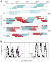
(A) Circadian rhythms of neuronal firing recorded from two SCN neurons in dispersed culture over a period of four weeks, showing that the cells oscillate independently with distinct circadian periods. Colored bars represent firing rates above the daily mean for the two cells (blue and red), with time of day plotted left to right and successive days top to bottom. The oscillations continue with unaltered phases after blockade of neuronal firing for 2.5 days by TTX (marked at right). (B) Circadian firing rhythm of one cell before and after TTX. Reprinted from Welsh et al. (7), with permission from Elsevier.
Figure 3
Figure 3
(A) Schematic diagram of intercellular signaling mechanisms within SCN, showing that action potentials arriving at synaptic terminals of core neurons trigger release of GABA and colocalized neuropeptides such as VIP or GRP. VIP, for example, binds to postsynaptic VPAC2 receptors of shell neurons, leading to membrane depolarization, elevated calcium and cAMP levels, phosphorylation of CREB, and induction of Per and Cry transcription, thus altering the phase and amplitude of the intracellular oscillator. Adjacent SCN neurons are also coupled by gap junctions. (B) The SCN network synchronizes and reinforces cellular oscillations, increases precision, and rescues rhythmicity of mutant oscillators. Reprinted from Liu et al. (209), with permission from McMillan Publishers Ltd.
Figure 4
Figure 4
Patterns of PER2::LUC bioluminescence recorded from mutant Cry1−/− neurons in a cultured slice (A, C) or in dispersed culture (B, D) over a period of six days. Bioluminescence images are shown above (A, B), from indicated times in hours after start of the experiment. Raster plots are shown below (C, D), with each horizontal line representing one cell, and values above and below the mean shown in red and green, respectively. Cry1−/− neurons are rhythmic and synchronized in SCN slices, but are not rhythmic in dispersed culture. Figure from Liu et al. (17), with permission from Elsevier.

Similar articles

Cited by

References

    1. Lowrey PL, Takahashi JS. Mammalian circadian biology: elucidating genome-wide levels of temporal organization. Annu Rev Genomics Hum Gen. 2004;5:407–441. - PMC - PubMed
    1. Ueda HR, et al. System-level identification of transcriptional circuits underlying mammalian circadian clocks. Nat Genet. 2005;37:187–92. - PubMed
    1. Yan J, Wang H, Liu Y, Shao C. Analysis of gene regulatory networks in the mammalian circadian rhythm. PLoS Comput Biol. 2008;4:e1000193. - PMC - PubMed
    1. Takahashi JS, Hong HK, Ko CH, McDearmon EL. The genetics of mammalian circadian order and disorder: implications for physiology and disease. Nat Rev Genet. 2008;9:764–75. - PMC - PubMed
    1. Klein DC, Moore RY, Reppert SM, editors. Suprachiasmatic Nucleus: The Mind’s Clock. New York: Oxford University Press; 1991.

Publication types

LinkOut - more resources