Review
. 2010:72:605-24.
doi: 10.1146/annurev-physiol-021909-135815.

Circadian organization of behavior and physiology in Drosophila

Affiliations
Review

Circadian organization of behavior and physiology in Drosophila

Ravi Allada et al. Annu Rev Physiol. 2010.

Abstract

Circadian clocks organize behavior and physiology to adapt to daily environmental cycles. Genetic approaches in the fruit fly, Drosophila melanogaster, have revealed widely conserved molecular gears of these 24-h timers. Yet much less is known about how these cell-autonomous clocks confer temporal information to modulate cellular functions. Here we discuss our current knowledge of circadian clock function in Drosophila, providing an overview of the molecular underpinnings of circadian clocks. We then describe the neural network important for circadian rhythms of locomotor activity, including how these molecular clocks might influence neuronal function. Finally, we address a range of behaviors and physiological systems regulated by circadian clocks, including discussion of specific peripheral oscillators and key molecular effectors where they have been described. These studies reveal a remarkable complexity to circadian pathways in this "simple" model organism.


Figures
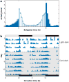
Figure 1
Figure 1
Drosophila circadian locomotor behavior. (a) An averaged locomotor activity plot for wild-type flies. Light and dark bars indicate normalized activity levels during 12 h of light and 12 h of dark, respectively. Increases in activity are evident in advance of light-to-dark and dark-to-light transitions. (b) An activity plot, or actogram, for a single wild-type fly. The height of vertical bars indicates level of activity during a 30-min interval, or bin. Each horizontal line contains 48 h of activity data (double-plotted plot), with the second day of data on one line repeated on the first day of data on the following line to ease visualization of the circadian period. The transition from light-dark conditions (LD) to dark-dark conditions (DD) has been indicated by a dashed horizontal line. Under DD, activity bouts occur at the same time each day, indicating a free-running period of near 24 h.
Figure 2
Figure 2
The core molecular clock in Drosophila. CLOCK/CYCLE (CLK/CYC) bind to E-box elements (E) contained in the promoters of period ( per) and timeless (tim). PER and TIM proteins are modified by the kinases DOUBLETIME (DBT), CASEIN KINASE 2 (CK2), and SHAGGY (SGG) and the phosphatases PROTEIN PHOSPHATASE 2A (PP2A) and PROTEIN PHOSPHATASE 1 (PP1). PER and TIM dimerize and transition to the nucleus, where they repress CLK/CYC activity. Phosphorylated PER and TIM also bind the E3 ubiquitin ligase SUPERNUMERARY LIMBS (SLIMB), which leads to ubiquitination and ultimately proteolysis by the 26S proteasome.
Figure 3
Figure 3
Additional feedback loops in the Drosophila molecular clockwork. In addition to the principal PERIOD/TIMELESS (PER/TIM) feedback loop, CLOCK/CYCLE (CLK/CYC) also activate CLOCKWORK ORANGE (CWO), PAR DOMAIN PROTEIN 1 (PDP1), and VRILLE (VRI). CWO feeds back to repress CLK/CYC activation by binding to E-box elements (E). PDP1 activates the transcription of the Clk gene, whereas VRI competes with PDP1 binding to repress Clk expression. P/V domain denotes PDP/VRI-binding sites.
Figure 4
Figure 4
The circadian pacemaker network in adult Drosophila brains. (a) Immunofluorescence of the circadian neuropeptide PIGMENT-DISPERSING FACTOR (PDF) illustrates the divergent projections stemming from ventral lateral neurons to other neural loci in the central nervous system. The small ventral lateral neurons (sLNv) send projections dorsally (toward the top of the image), whereas the large ventral lateral neurons (lLNv) send projections contralaterally and extensively into the optic lobes (on the right and left of the image). (b) tim in-situ hybridizations reveal the spatial orientation of pacemaker neurons in the central nervous system. Labels denote the location of the ventral lateral neuron (LNv), dorsal lateral neuron (LNd), and dorsal neuron (DN) clusters. Images in both panels a and b were provided by Ela Kula-Eversole. (c) Schematic of the Drosophila circadian pacemaker network. sLNv (orange) send projections dorsally. lLNv (maroon) send projections contralaterally and into the optic lobes (left and right sides of the schematic). LNd are indicated in yellow, and three groups of dorsal neurons are indicated in green.

Similar articles

Cited by

References

    1. Takahashi JS, Hong HK, Ko CH, McDearmon EL. The genetics of mammalian circadian order and disorder: implications for physiology and disease. Nat Rev Genet. 2008;9:764–75. - PMC - PubMed
    1. Pittendrigh C. On temperature independence in the clock-system controlling emergence time in Drosophila. Proc Natl Acad Sci USA. 1954;40:1018–29. - PMC - PubMed
    1. Dubruille R, Emery P. A plastic clock: how circadian rhythms respond to environmental cues in Drosophila. Mol Neurobiol. 2008;38:129–45. - PubMed
    1. Nitabach MN, Taghert PH. Organization of the Drosophila circadian control circuit. Curr Biol. 2008;18:R84–93. - PubMed
    1. Zheng X, Sehgal A. Probing the relative importance of molecular oscillations in the circadian clock. Genetics. 2008;178:1147–55. - PMC - PubMed

Publication types

LinkOut - more resources