Randomized Controlled Trial
. 2010 Sep;65(9):1126-33.
doi: 10.1111/j.1398-9995.2010.02327.x. Epub 2010 Feb 10.

CC chemokine receptor 4 (CCR4) in human allergen-induced late nasal responses

Affiliations
Randomized Controlled Trial

CC chemokine receptor 4 (CCR4) in human allergen-induced late nasal responses

G Banfield et al. Allergy. 2010 Sep.

Abstract

Background: CC Chemokine receptor 4 (CCR4) is preferentially expressed on Th2 lymphocytes. CCR4-mediated inflammation may be important in the pathology of allergic rhinitis. Disruption of CCR4 - ligand interaction may abrogate allergen-induced inflammation.

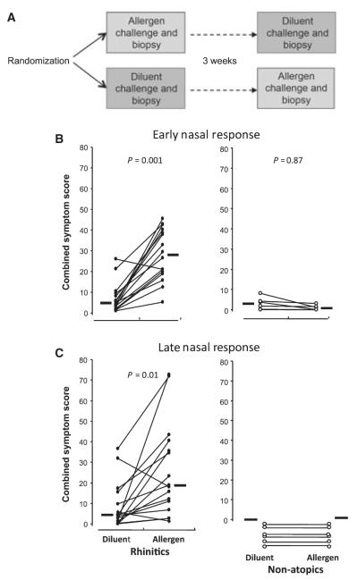
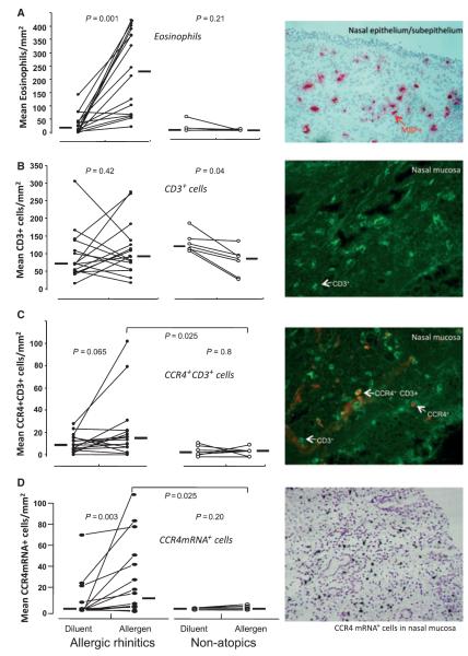
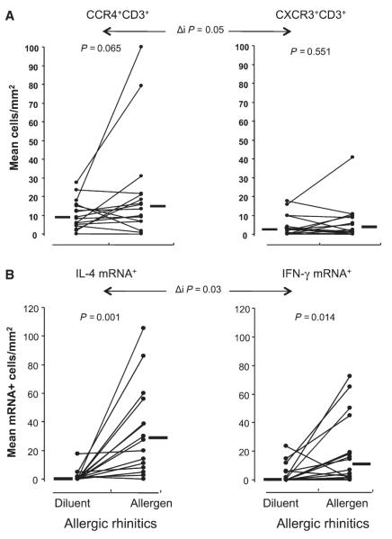
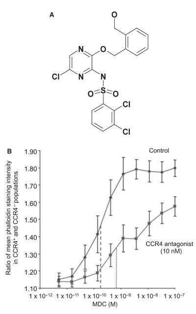
Methods: Sixteen allergic rhinitics and six nonatopic individuals underwent both allergen and control (diluent) nasal challenges. Symptom scores and peak nasal inspiratory flow were recorded. Nasal biopsies were taken at 8 h post challenge. Sections were immunostained and examined by light or dual immunofluorescence microscopy for eosinophils, T-lymphocytes, CCR4(+)CD3(+) and CXCR3(+)CD3(+) cells and examined by in situ hybridization for CCR4, IL-4 and IFN-gamma mRNA(+) cells. Peripheral blood mononuclear cells were obtained from peripheral blood of nine normal donors and the CCR4(+)CD4(+) cells assessed for actin polymerization in response to the CCR4 ligand macrophage-derived chemokine (MDC/CCL22) and the influence of a CCR4 antagonist tested.

Results: Allergic rhinitics had increased early and late phase symptoms after allergen challenge compared to diluent; nonatopics did not respond to either challenge. Eosinophils, but not total numbers of CD3(+) T cells, were increased in rhinitics following allergen challenge. In rhinitics, there was an increase in CCR4(+)CD3(+) protein-positive cells relative to CXCR3(+)CD3(+) cells; CCR4 mRNA+ cells were increased and IL-4 increased to a greater extent than IFN-gamma. CCR4(+)CD4(+) T cells responded to MDC in vitro, and this response was inhibited by the selective CCR4 antagonist.

Conclusion: Lymphocyte CCR4 expression is closely associated with induction of human allergen-induced late nasal responses. Blocking CCR4-ligand interaction may provide a novel therapeutic approach in allergic disease.


Figures

Figure 1
Figure 1
(A) Participants were randomized to one of two nasal challenge protocols: either undergoing allergen challenge to one nostril selected at random plus biopsy, followed by diluent challenge to the other nostril 3 weeks later, or vice versa. (B) Early phase response to intranasal challenge with 2500 BU allergen as nasal spray or equal volume of diluent alone. Symptom scores for nasal itch/sneezing, running and blockage were recorded on a visual analogue scale at 15, 30 and 60 min and plotted on a graph. The area under the curve was then calculated and expressed as the early phase combined symptom score. (C) Late phase response to nasal allergen or diluent challenge. Symptom scores were recorded at hourly intervals; the combined symptom score was calculated as the area under the curve between 2 and 8 h. Circles represent individual participants; lines connect responses in the same individual. Horizontal bars represent median values. P-values represent comparison by Wilcoxon matched-pairs sign rank test.
Figure 2
Figure 2
(A) left; mean number of eosinophils per square millimeter counted by light microscopy in nasal mucosal biopsy specimens taken from atopic and nonatopic participants at 8 h after intranasal challenge with diluent or allergen, right; example of eosinophils as identified by positive immunostaining for major basic protein (MBP) in nasal mucosa (× 200 magnification). (B) left; mean number of CD3+ cells per square millimeter counted by fluorescent microscopy following immunostaining in nasal biopsy specimens, right; example of staining. (C) left; mean number of CC Chemokine receptor 4 (CCR4)+CD3+ cells per square millimeter in the nasal mucosa, right; example of individual and dual CCR4+CD3+ cells following dual immunohistochemistry identified by fluorescence microscopy (× 400 magnification). (D) left; mean number of CCR4 mRNA+ cells per square millimeter following in situ hybridization in nasal mucosa specimens, right; example of CCR4 mRNA positive cells identified by dense accumulation of silver grains overlying individual cells (× 200 magnification). Circles represent individual participants; lines connect measurements in the same individual. Horizontal bars represent median values. P-values represent within-group comparison by Wilcoxon matched-pairs sign rank test or between-group comparison by Mann–Whitney U-test.
Figure 3
Figure 3
(A) mean numbers of CC Chemokine receptor 4+CD3+ (left) and CXCR3+CD3+ cells (right) as identified by dual immunofluorescence in the nasal mucosa at 8 h after diluent or allergen challenge in atopic individuals only. (B) mean number of IL-4 (left) and IFN-γ (right) mRNA+ cells per square millimeter following in situ hybridization of nasal mucosa specimens from atopic participants after diluent or allergen challenge. Circles represent individual participants; lines connect measurements in the same individual. Horizontal bars represent median values. P-values represent comparison by Wilcoxon matched-pairs sign rank test, Δi P-values represent comparison of degree of change in median number of positive cells between diluent and allergen challenge between groups by Mann–Whitney U-test.
Figure 4
Figure 4
(A) Structure of the selective CC Chemokine receptor 4 (CCR4) antagonist used in this study. (B) Ratio of mean phalloidin-staining intensity between CD4+CCR4+ and CD4+CCR4− cells measured following stimulation with increasing concentrations of the CCR4 agonist macrophage-derived chemokine (MDC) after prior incubation with CCR4 antagonist or buffer alone. Fluorescence intensity of 1000 CD4+CCR4+ cells following cell lysis and staining with FITC-phalloidin was normalized to mean intensity of CD4+CCR4− cells in the same sample. Individual data points represent the mean of nine experiments, bars represent standard error. Approximate EC50 values are plotted without (- - - -) and with (· · · ·) the addition of the CCR4 antagonist.

Similar articles

Cited by

References

    1. Cosmi L, Annunziato F, Maggi E, Romagnani S, Manetti R. Chemoattractant receptors expressed on type 2 T cells and their role in disease. Int Arch Allergy Immunol. 2001;125:273–279. - PubMed
    1. Bonecchi R, Bianchi G, Bordignon PP, D’Ambrosio D, Lang R, Borsatti A, et al. Differential expression of chemokine receptors and chemotactic responsiveness of type 1 T helper cells (Th1s) and Th2s. J Exp Med. 1998;187:129–134. - PMC - PubMed
    1. D’Ambrosio D, Iellem A, Bonecchi R, Mazzeo D, Sozzani S, Mantovani A, et al. Selective up-regulation of chemokine receptors CCR4 and CCR8 upon activation of polarized human type 2 Th cells. J Immunol. 1998;161:5111–5115. - PubMed
    1. Lloyd CM, Delaney T, Nguyen T, Tian J, Martinez-A C, Coyle AJ, et al. CC chemokine receptor (CCR3)/eotaxin is followed by CCR4/monocyte-derived chemokine in mediating pulmonary T helper lymphocyte type 2 recruitment after serial antigen challenge in vivo. J Exp Med. 2000;191:265–274. - PMC - PubMed
    1. Lloyd CM, Rankin SM. Chemokines in allergic airway disease. Curr Opin Pharmacol. 2003;3:443–448. - PMC - PubMed

Publication types

MeSH terms