. 2010 Mar;159(5):1092-105.
doi: 10.1111/j.1476-5381.2009.00633.x. Epub 2010 Feb 8.

Distinct expression and ligand-binding profiles of two constitutively active GPR17 splice variants

Affiliations

Distinct expression and ligand-binding profiles of two constitutively active GPR17 splice variants

T Benned-Jensen et al. Br J Pharmacol. 2010 Mar.

Abstract

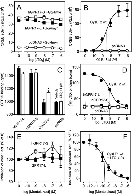
Background and purpose: In humans and non-human primates, the 7TM receptor GPR17 exists in two isoforms differing only by the length of the N-terminus. Of these, only the short isoform has previously been characterized. Hence, we investigated gene expression and ligand-binding profiles of both splice variants and furthermore uncovered and characterized constitutive activity of both isoforms.

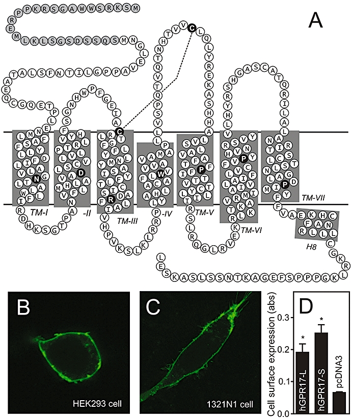
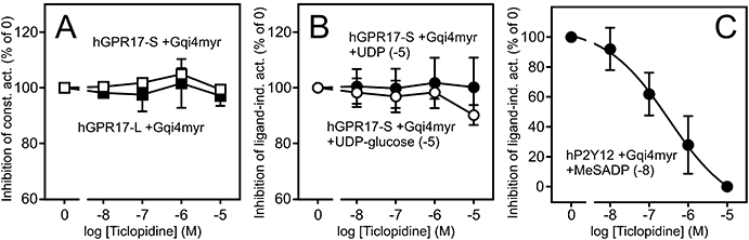
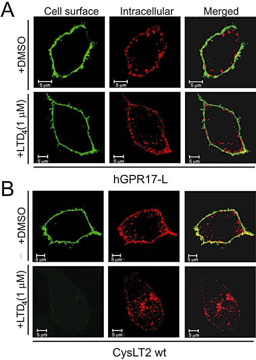
Experimental approach: Expression levels of the hGPR17 isoforms were determined in several brain regions as well as heart and kidney using quantitative RT-PCR. A CREB reporter assay and [(35)S]-GTPgammaS binding were employed to assess the constitutive activity and the activation by UDP, UDP-glucose and -galactose and the cysteinyl leukotrienes LTC(4) and LTD(4). Leukotriene binding and induction of internalization were furthermore tested using homologous competition binding and antibody-feeding experiments respectively.

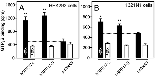
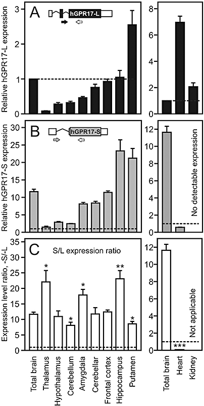
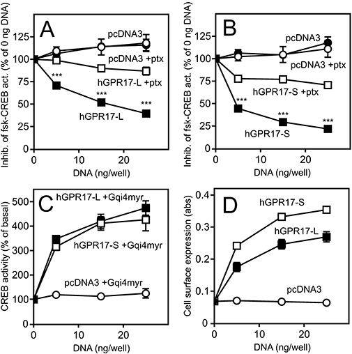
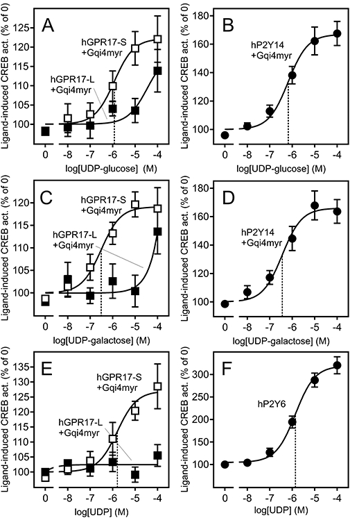
Key results: The short isoform (hGPR17-S) was expressed more abundantly (eight- to 23-fold) in the brain than the long isoform (hGPR17-L), whereas the opposite was observed in heart and kidney. As previously reported, the uracil nucleotides activated hGPR17-S with micromolar potencies. However, much lower potencies were observed for hGPR17-L with a 50- to 170-fold increase in EC(50). Furthermore, contrary to previous reports, neither of the isoforms was activated or bound by the cysteinyl leukotrienes. Finally, both receptors were demonstrated to be constitutively active through Galpha(i).

Conclusions and implications: We present the first isoform-specific characterization of GPR17 and show that differences exist between the isoforms, in both expression pattern and pharmacological profile. In turn, our results indicate that the two human isoforms might serve tissue-specific functions.


Figures

Figure 7
Figure 7
LTD4 does not activate or bind to either of the hGPR17 isoforms. (A) Response to LTD4 in HEK293 cells transiently transfected with Gqi4myr, CREB-Luc reporter plasmid and FLAG-tagged hGPR17-L, -S or pcDNA3 at 15 ng per well. The results represent mean ± SEM of raw data from four independent experiments performed in quadruplicates. (B) Activation of FLAG-tagged CysLT2 by LTD4; performed and presented as in (A). The EC50 value is indicated by the stippled line. (C) Effects of LTD4 1 µM (open columns) or vehicle (DMSO, solid columns) on [35S]-GTPγS binding to membranes isolated from HEK293 cells transiently transfected with FLAG-tagged hGPR17-L, -S, CysLT2 or pcDNA. The data represent mean ± SEM of raw data from four independent experiments performed in triplicates. *P<0.05 by Student's t-test. (D) Homologous competition assay for [3H]-LTD4 binding to the membranes from HEK293 cells transiently transfected with hGPR17-L, -S, CysLT2 or pcDNA3; these were similar to those used in (C). The data represent mean ± SEM of raw data from four independent experiments performed in duplicates. ipThe IC50 value for CysLT2 is indicated by the stapled line. (E) Putative inverse agonism of Montelukast. HEK293 cells were transfected as in (A) and incubated with Montelukast in varying concentrations. The results represent mean ± SEM of three independent experiments performed in triplicates and are given as % relative to the activity in absence of the ligand. (F) CysLT1 control. HEK293 cells were transfected as described in (A) and incubated with LTC4 at 1 nM and Montelukast at varying concentrations. The EC50 value is indicated by the stippled line. CREB, cAMP response element binding protein; hGPR17-L, long GPR17 isoform; hGPR17-S, short GPR17 isoform.
Figure 4
Figure 4
Constitutive signalling of hGPR17-L and -S as measured by [35S]-GTPγS binding. (A) [35S]-GTPγS binding to membranes isolated from HEK293 cells transiently transfected with FLAG-tagged hGPR17-L, -S or pcDNA3. The cells were cultured in the absence (solid columns) or presence (open columns) of 100 ng·mL–1 pertussis toxin (ptx). The level of binding to pcDNA3-membranes is indicated by the stippled line. The results represent mean ± SEM of raw data from four independent experiments performed in triplicates. **P<0.01 by Student's t-test. (B) [35S]-GTPγS binding to membranes isolated from 1321N1 cells transiently transfected with hGPR17-L, -S or pcDNA3; performed and presented as in (A). *P<0.05, **P<0.01 by Student's t-test. hGPR17-L, long GPR17 isoform; hGPR17-S, short GPR17 isoform.
Figure 2
Figure 2
Gene expression levels of the two hGPR17 isoforms and the ratio of these in brain, heart and kidney. (A) Levels of hGPR17-L transcript in total brain and eight different brain regions (left panel) and kidney and heart (right panel). The expression levels are given relative (in fold) to that in total brain, which is indicated by the stippled line. The insert is a schematic representation of the hGPR17-L mRNA species. Translated and untranslated regions are indicated in black and white respectively. The isoform-specific forward primer spans the exon 2–3 border and is depicted in black, whereas the shared reverse primer is depicted in white. (B) Levels of hGPR17-S the same tissues as in (A). The expression levels are given relative (in fold) to that of hGPR17-L in total brain (stippled line). No expression was detected in kidney. The insert is a schematic representation of the hGPR17-S mRNA species. Translated and untranslated regions are indicated in grey and white respectively. The isoform-specific forward primer spans the exon 1–3 border and is depicted in grey, whereas the shared reverse primer is depicted in white. (C) Ratio of hGPR17-S and -L expression levels from (B) and (A) respectively. The stippled line indicates a ratio of 1, i.e. equal expression levels. Ratios significantly different from that in total brain are indicated; *P<0.05, **P<0.01 and ***P<0.0001 (Student's t-test). Given the absence of hGPR17-S expression in kidney the ratio is not calculable in this instance. hGPR17-L, long GPR17 isoform; hGPR17-S, short GPR17 isoform.
Figure 1
Figure 1
Primary structure and cell surface expression of hGPR17. (A) Serpentine model of hGPR17. The highly conserved residues among rhodopsin-like 7TM receptors are indicated as black circles with white letters in each transmembrane helix and the conserved disulphide-bridge between the extracellular loop 2 and the conserved cysteine residue in TM3 denoted as a stapled line. The 28 amino acids comprising the longer N-terminus of the hGPR17-L splice variant are indicated as grey circles. (B) Expression of C-terminally eGFP-tagged hGPR17-L in transiently transfected HEK293 cells as detected by confocal microscopy. (C) Expression of C-terminally eGFP-tagged hGPR17-L in transiently transfected 1321N1 cells as detected by confocal microscopy. (D) Cell surface expression of FLAG-tagged hGPR17 isoforms in HEK293 cells transiently transfected with hGPR17-L, -S or pcDNA3 vector control as measured by ELISA. The results represent mean ± SEM of raw data from three independent experiments performed in quadruplicates. *P<0.05 by Student's t-test. 7TM, seven transmembrane; hGPR17-L, long GPR17 isoform.
Figure 3
Figure 3
Constitutive signalling (A–C) and surface expression (D) of the two hGPR17 isoforms. (A) Inhibition of forskolin-induced CREB activity (abbreviated fsk-CREB act.) in HEK293 cells transfected with hGPR17-L or pcDNA3 vector control in the presence or absence of pertussis toxin (ptx, 100 ng·mL–1). The cells were transiently transfected with CREB-Luc reporter vector and FLAG-tagged hGPR17-L or pcDNA3 at 0, 5, 15 and 25 ng per well. Production of cAMP was stimulated with 15 µM forskolin. The results are given relative to the activity at 0 ng per well in % and are presented as mean ± SEM from at least four independent experiments performed in quadruplicates. (B) Inhibition of forskolin-induced cAMP production in HEK293 cells transfected with FLAG-tagged hGPR17-S or pcDNA3; performed and presented as in (A). Statistical analyses of data in (A) and (B): ***P<0.0001 (Student's t-test). (C) CREB activity in HEK293 cells transiently transfected with the chimeric Gα subunit Gqi4myr, CREB-Luc reporter vector and hGPR17-L, -S or pcDNA3 at 0, 5, 15 and 25 ng per well. The results are given relative to the activity at 0 ng per well in % and are presented as mean ± SEM from four independent experiments performed in quadruplicates. (D) Cell surface expression of FLAG-tagged hGPR17-L and -S in HEK293 cells as measured by ELISA. The cells were transiently transfected with hGPR17-L, -S or pcDNA3 in parallel with the experiments presented in (C). The results represent mean ± SEM of raw data from four independent experiments performed in quadruplicates. CREB, cAMP response element binding protein; hGPR17-L, long GPR17 isoform; hGPR17-S, short GPR17 isoform.
Figure 5
Figure 5
Uracil nucleotide ligands activate hGPR17-L and -S with different potencies. (A) Activation of FLAG-tagged hGPR17-L and -S by UDP-glucose in transiently transfected HEK293 cells. The cells were transfected with Gqi4myr, CREB-Luc reporter plasmid and receptor or pcDNA3 at 15 ng per well. The results are corrected for background (pcDNA3-transfected cells) and given as % relative to the activity in absence of the ligand. The data represent mean ± SEM of four independent experiments performed in quadruplicates. The EC50 of the hGPR17-S response is indicated by the stippled line. (B) Activation of hP2Y14 by UDP-glucose in transiently transfected HEK293 cells; performed and presented as in (A). (C) Activation of FLAG-tagged hGPR17-L and -S by UDP-galactose in transiently transfected HEK293 cells; performed and presented as in (A). (D) Activation of hP2Y14 by UDP-galactose in transiently transfected HEK293 cells; performed and presented as in (A). (E) Activation of FLAG-tagged hGPR17-L and -S by UDP in transiently transfected HEK293 cells; performed and presented as in (A). (F) Activation of hP2Y6 by UDP in transiently transfected HEK293 cells; performed and presented as in (A) with the exception that Gqi4myr was not co-transfected in this case. CREB, cAMP response element binding protein; hGPR17-L, long GPR17 isoform; hGPR17-S, short GPR17 isoform.
Figure 6
Figure 6
Ticlopidine does not display inverse agonism or antagonism at hGPR17 isoforms. (A) Putative inverse agonism of Ticlopidine. HEK293 cells were transiently transfected with Gqi4myr, CREB-Luc reporter plasmid and receptor or pcDNA3 at 15 ng per well.and incubated with the ligand in varying concentrations. The results are corrected for background (pcDNA3-transfected cells) and given as % relative to the activity in absence of the ligand. The data represent mean ± SEM of two independent experiments performed in quadruplicates. (B) Putative antagonism of Ticlopidine. HEK293 cells were transfected as described in (A) and incubated with agonist (UDP or UDP-glucose) at 10 µM and Ticlopidine in varying concentrations. The results are presented as in (A). (C) hP2Y12 control. HEK293 cells were transfected as described in (A) and were incubated with the agonist MeSADP at 10 nM and Ticlopidine in varying concentrations. The results are presented as in (A). CREB, cAMP response element binding protein; hGPR17, GPR17 isoform.
Figure 8
Figure 8
LTD4 does not induce internalization of hGPR17-L. (A) Internalization of FLAG-tagged hGPR17-L in transiently transfected HEK293 cells in the presence of vehicle (DMSO, upper panels) or LTD4 at 1 µM (lower panels). Receptors at the surface were labelled with M1 anti-FLAG antibody prior to internalization. Subsequent to this, labelled receptors still residing at the cell surface were detected before permeabilization (green, left) and internalized receptors after (red, middle) with two different secondary antibodies and analysed by confocal microscopy. Merged images are presented to the right. (B) Internalization of FLAG-tagged CysLT2 in the presence of vehicle or LTD4; performed and presented as in (A). Scale bars denote 5 µM. hGPR17-L, long GPR17 isoform.

Similar articles

Cited by

References

    1. Alexander SP, Mathie A, Peters JA. Guide to Receptors and Channels (GRAC), 3rd edition. Br J Pharmacol. 2008;153(Suppl 2):S1–S209. - PMC - PubMed
    1. Benned-Jensen T, Rosenkilde MM. Structural motifs of importance for the constitutive activity of the orphan 7TM receptor EBI2: analysis of receptor activation in the absence of an agonist. Mol Pharmacol. 2008;74:1008–1021. - PubMed
    1. Blasius R, Weber RG, Lichter P, Ogilvie A. A novel orphan G protein-coupled receptor primarily expressed in the brain is localized on human chromosomal band 2q21. J Neurochem. 1998;70:1357–1365. - PubMed
    1. Ceruti S, Villa G, Genovese T, Mazzon E, Longhi R, Rosa P, et al. The P2Y-like receptor GPR17 as a sensor of damage and a new potential target in spinal cord injury. Brain. 2009;132:2206–2218. - PubMed
    1. Chambers JK, Macdonald LE, Sarau HM, Ames RS, Freeman K, Foley JJ, et al. A G protein-coupled receptor for UDP-glucose. J Biol Chem. 2000;275:10767–10771. - PubMed

Publication types