. 2010 Apr;23(2):190-200.
doi: 10.1111/j.1755-148X.2010.00685.x. Epub 2010 Feb 10.

PLX4032, a selective BRAF(V600E) kinase inhibitor, activates the ERK pathway and enhances cell migration and proliferation of BRAF melanoma cells

Affiliations
Free PMC article

PLX4032, a selective BRAF(V600E) kinase inhibitor, activates the ERK pathway and enhances cell migration and proliferation of BRAF melanoma cells

Ruth Halaban et al. Pigment Cell Melanoma Res. 2010 Apr.
Free PMC article

Erratum in

  • Pigment Cell Melanoma Res. 2012 May;25(3):402

Abstract

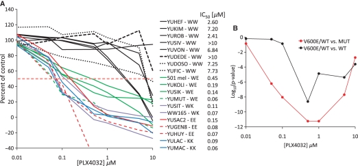
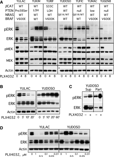
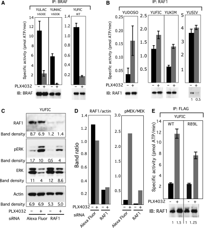
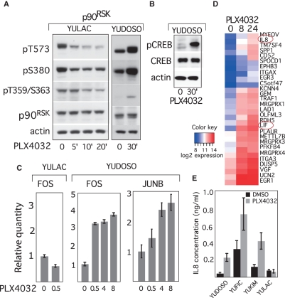
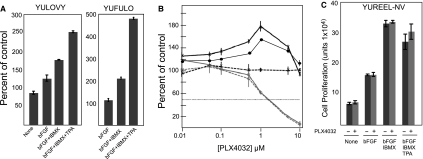
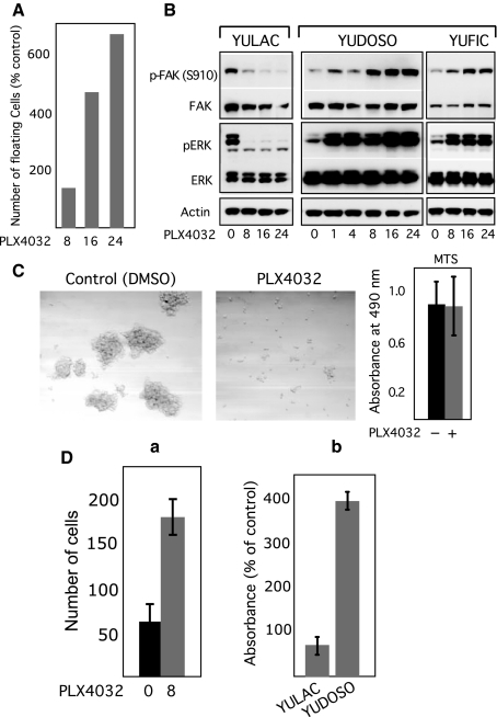
BRAF(V600E/K) is a frequent mutationally active tumor-specific kinase in melanomas that is currently targeted for therapy by the specific inhibitor PLX4032. Our studies with melanoma tumor cells that are BRAF(V600E/K) and BRAF(WT) showed that, paradoxically, while PLX4032 inhibited ERK1/2 in the highly sensitive BRAF(V600E/K), it activated the pathway in the resistant BRAF(WT) cells, via RAF1 activation, regardless of the status of mutations in NRAS or PTEN. The persistently active ERK1/2 triggered downstream effectors in BRAF(WT) melanoma cells and induced changes in the expression of a wide-spectrum of genes associated with cell cycle control. Furthermore, PLX4032 increased the rate of proliferation of growth factor-dependent NRAS Q61L mutant primary melanoma cells, reduced cell adherence and increased mobility of cells from advanced lesions. The results suggest that the drug can confer an advantage to BRAF(WT) primary and metastatic tumor cells in vivo and provide markers for monitoring clinical responses.


Figures

Figure 1
Figure 1
Growth responses to PLX4032. (A) CellTiter-Glo® Luminescent Cell Viability Assay. Values are percent of control (DMSO) assessed at the end of 72-h treatment with PLX4032 (1 μM). Each measurement is the average of triplicate or quadruplet wells. The legend on the right provides the IC50 indicating melanoma cells that are BRAF homozygous or heterozygous for wild-type (W), V600K (K) or V600E (E) mutation. Among the BRAFWT cell strains, YUDOSO and YUFIC are heterozygous for the NRAS Q61K and Q61R mutation, respectively (dotted black line), and YUDEDE cells are null for PTEN and carry homozygous NRAS Q61H mutation (dashed black line). Green and red dashed lines indicate BRAF mutant cells null for PTEN. The horizontal dashed line marked IC50. STDV was about 5% of total count. (B) A two-sample Wilcoxon rank sum test assessing differences in growth responses of wild-type and mutant melanoma cells at several concentrations of PLX4032. The graphs, plotted as logarithm of the P-values (y-axis) over PLX4032 concentration show significant differences over a broad range of PLX4032 concentrations in the response of cells carrying the heterozygous BRAFV600E/WT alleles compared to BRAFWT melanoma cells (red), and BRAFV600E/WT compared with other mutant cells, i.e. homozygous BRAFV600E/E, BRAFV600K/K or heterozygous BRAFV600K/WT (black).
Figure 2
Figure 2
Changes in ERK1/2 and MEK in response to PLX4032. (A) Melanoma cell strains were treated with DMSO for 4 h (0), or with PLX4032 (1 μM) for 4 and 8 h. The panels show Western blots probed with antibodies to phosph-ERK1/2 Thr202/Tyr204 mAb (pERK), ERK1/2 (ERK), phospho-MEK1/2 (pMEK), MEK1/2 (MEK), and actin as a loading control. The mutation status of BRAF, NRAS, PTEN and β-catenin (βCAT) are indicated at the top. (B) Western blot analyses of ERK1/2 inactivation/activation after short-term incubation with PLX4032 (1 μM). (C) pERK1/2 and ERK1/2 in supernatant (Sup) and particulate (Part) fractions. (D) Changes in pERK1/2 activation after treatments with increasing concentration of PLX4032, or DMSO for 1 h.
Figure 3
Figure 3
RAF kinase in response to PLX4032. (A, B, E) Immune-complex kinase activities expressed as picomoles [γ-32P]-ATP incorporated into MBP. Each data point is an average of triplicate measurements ± STDV. The cells were treated with PLX4032 for 1 h (A), 4 h (B) and 16 h (E). The panels under each kinase assay shows Western blots of immunoprecipitated proteins eluted with SDS sample buffer and probed with antibodies to the respective protein. Numbers under the protein bands in (B) and (E) indicate relative band density determined by densitometric analysis. (C) Suppression of PLX4032 induced ERK activation in YUFIC-BRAFWT melanoma after RAF1 depletion by siRNA. YUFIC melanoma cells were transfected with Alexa Fluor or RAF1 siRNA and 2 days later were treated with PLX4032 or DMSO for 1 h. Cell lysates were probed with the indicated antibodies. The numbers under each blot represent values of band density in pixels 1 × 10³. (D) Histograms showing band intensity ratios normalizing RAF1 to actin and pERK to total ERK as presented in (C).
Figure 4
Figure 4
Inactivation/activation of downstream ERK targets in response to PLX4032. (A, B) Western blots showing changes in p90RSK and CREB activation states, respectively, employing phospho-specific antibodies. (C) Changes in c-FOS and JUNB gene transcripts in response to PLX4032 as evaluated by Real Time RT-PCR. Data are averages of three replicates ± STDV. (D) Heatmap showing upregulation of gene expression by at least 3-fold and above in YUDOSO-BRAFWT melanoma cells in response to treatment with PLX4032 for 8 and 24 h employing the NimbleGen whole genome expression arrays. Marked in red circles are IL8 and LIF. (E) ELISA assay confirming an increase in secreted IL8 levels after 24 h incubation with PLX4032 in BRAFWT (YUDOSO, YUFIC and YUKIM), but not in mutant melanoma cells (YULAC). Error bars represent STDV of six wells.
Figure 5
Figure 5
Proliferation of mitogen-dependent cells in response to PLX4032. (A) YULOVY and YUFULO cell proliferation after incubation for 72 h in basal medium (none) or medium supplemented with growth factors as indicated. Values are averages of triplicate wells expressed as percent of control (Day 0). (B) Dose response to increasing concentrations of PLX4032. YULOVY and YUFULO, solid dark lines with X and solid circles, respectively, grown in medium supplemented with bFGF and IBMX; YULOVY cells grown with bFGF and IBMX, broken dark line; Keratinocytes grown without and with supplements, gray solid and broken lines, respectively. Other details as in Figure 1. (C) YUREEL-NV melanocyte proliferation in basal medium without growth factors (none), and with growth factors (as indicated) in the absence and presence of PLX4032. Error bars represent STDV of triplicate wells.
Figure 6
Figure 6
PLX4032 reduced cell adhesion and promoted migration in BRAFWT melanoma cells. (A) The histograms show the number of floating YUDOSO-BRAFWT melanoma cells at different time intervals (in hours) after the addition of PLX4032 as percent of control (2500 floating cells). (B) Changes in FAK activation in response to PLX4032, as detected by Western blotting with antibodies against phospho FAK (S910), FAK, phospho-ERK1/2, ERK1/2 and actin. (C) Low magnification photomicrographs of 10 days soft agar cultures showing YUDOSO-BRAFWT cells forming large colonies in control (DMSO), but single cells or small colonies in PLX4032. The histogram demonstrates the viability (MTS) of cells in 96-well soft agar plates measuring absorbance at 490 nm. (D) Cell migration in response to PLX4032. The histograms show the number of cells that migrated thorough the transwell pores by counting three microscopical fields after 8 h treatment (a), or by extracting the stained cells and measuring absorbance at 570 nm after 24 h incubation with the drug normalized to control, DMSO treated cells (b).

References

    1. Baccarini M. Second nature: biological functions of the Raf-1 “kinase”. FEBS Lett. 2005;579:3271–3277. - PubMed
    1. Bild AH, Yao G, Chang JT, et al. Oncogenic pathway signatures in human cancers as a guide to targeted therapies. Nature. 2006;439:353–357. - PubMed
    1. Böhm M, Moellmann G, Cheng E, Alvarez-Franco M, Wagner S, Sassone-Corsi P, Halaban R. Identification of p90RSK as the probable CREB-Ser133 kinase in human melanocytes. Cell Growth Differ. 1995;6:291–302. - PubMed
    1. Craig EA, Stevens MV, Vaillancourt RR, Camenisch TD. MAP3Ks as central regulators of cell fate during development. Dev. Dyn. 2008;237:3102–3114. - PubMed
    1. Curtin JA, Busam K, Pinkel D, Bastian BC. Somatic activation of KIT in distinct subtypes of melanoma. J. Clin. Oncol. 2006;24:4340–4346. - PubMed

Publication types

MeSH terms