. 2010 Feb 11:7:36.
doi: 10.1186/1743-422X-7-36.

Impaired antiviral activity of interferon alpha against hepatitis C virus 2a in Huh-7 cells with a defective Jak-Stat pathway

Affiliations

Impaired antiviral activity of interferon alpha against hepatitis C virus 2a in Huh-7 cells with a defective Jak-Stat pathway

Sidhartha Hazari et al. Virol J. .

Abstract

Background: The sustained virological response to interferon-alpha (IFN-alpha) in individuals infected with hepatitis C virus (HCV) genotype 1 is only 50%, but is about 80% in patients infected with genotype 2-6 viruses. The molecular mechanisms explaining the differences in IFN-alpha responsiveness between HCV 1 and other genotypes have not been elucidated.

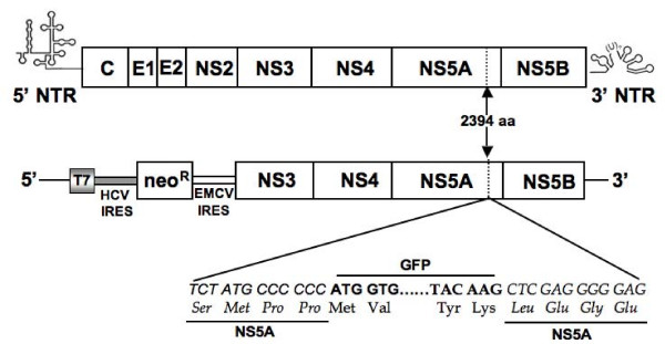
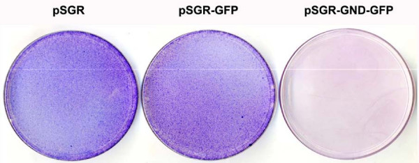
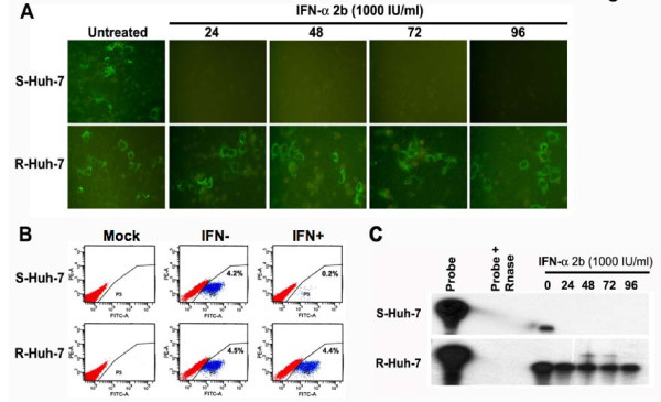
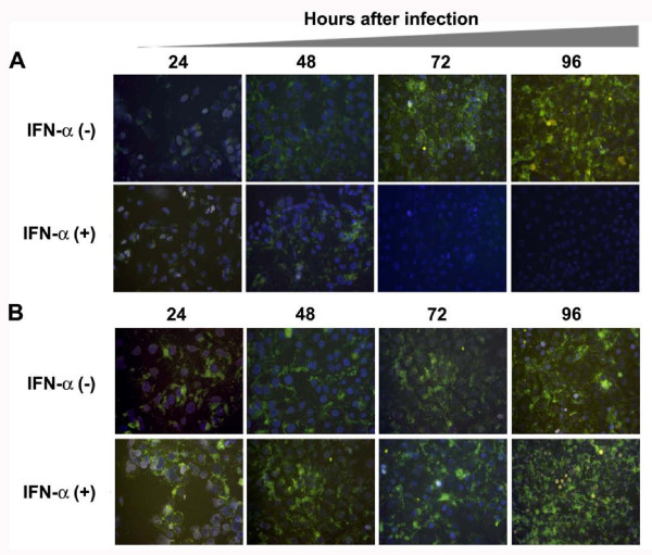
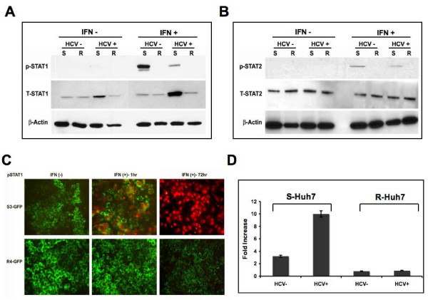
Results: Virus and host cellular factors contributing to IFN responsiveness were analyzed using a green fluorescence protein (GFP) based replication system of HCV 2a and Huh-7 cell clones that either possesses or lack a functional Jak-Stat pathway. The GFP gene was inserted into the C-terminal non-structural protein 5A of HCV 2a full-length and sub-genomic clones. Both HCV clones replicated to a high level in Huh-7 cells and could be visualized by either fluorescence microscopy or flow cytometric analysis. Huh-7 cells transfected with the GFP tagged HCV 2a genome produced infectious virus particles and the replication of fluorescence virus particles was demonstrated in naïve Huh-7.5 cells after infection. IFN-alpha effectively inhibited the replication of full-length as well as sub-genomic HCV 2a clones in Huh-7 cells with a functional Jak-Stat pathway. However, the antiviral effect of IFN-alpha against HCV 2a virus was not observed in Huh-7 cell clones with a defect in Jak-Stat signaling. HCV infection or replication did not alter IFN-alpha induced Stat phosphorylation or ISRE promoter-luciferase activity in both the sensitive and resistant Huh-7 cell clones.

Conclusions: The cellular Jak-Stat pathway is critical for a successful IFN-alpha antiviral response against HCV 2a. HCV infection or replication did not alter signaling by the Jak-Stat pathway. GFP labeled JFH1 2a replicon based stable cell lines with IFN sensitive and IFN resistant phenotypes can be used to develop new strategies to overcome IFN-resistance against hepatitis C.


Figures

Figure 1
Figure 1
Structure of HCV full-length and sub-genomic clones used in this project. The coding sequence of GFP was inserted in frame with the NS5A coding sequence of JFH1 cDNA clone between 2394 and 2395 amino acids position (between 417 and 418 amino acids of NS5A protein). Changes in the nucleotide and amino acid sequences of NS5A gene of HCV-GFP chimera clone are shown. GFP was also inserted at the similar location of NS5A gene (between 417 and 418 amino acids) in the sub-genomic clone, GND mutant clone and E1-E2 deleted mutant clone.
Figure 2
Figure 2
Replication of JFH1-GFP full-length RNA and JFH1-GND-GFP mutant RNA in Huh-7.5 cells after transfection. Huh-7.5 cells were electroporated with 10 μg of in vitro transcribed RNA prepared either from full-length or GND mutant plasmid. Intracellular expression of GFP in the transfected culture was examined under a fluorescence microscope. (A) Intracellular GFP expression in Huh-7.5 cells transfected with JFH1-GFP RNA at 0, 24, 48, 72 and 96 hours. (B) Intracellular expression of GFP in Huh-7.5 cells transfected with JFH1-GND-GFP mutant RNA at 0, 24, 48, 72 and 96 hours. (C) Intracellular GFP expression measured by flow cytometry in the Huh-7.5 cells transfected with JFH1-GFP RNA after 72 hours. (D): Intracellular GFP expression measured by flow cytometry in the transfected cells of JFH1-GND-GFP mutant RNA after 72 hours.
Figure 3
Figure 3
Detection of positive and negative strand HCV RNA in the transfected Huh 7.5 cells by RPA. Huh-7.5 cells were transfected with 10 μg of in vitro transcribed full-length JFH1-GFP and JFH1-GND-GFP mutant HCV RNA by electroporation. Total RNA was isolated from the RNA transfected cell culture at 0, 24, 48, 72 and 96 hours post-transfection. For the detection of positive strand HCV RNA, total cellular RNA was hybridized with a negative strand RNA probe targeted to the highly conserved 5'UTR region and then RPA experiment was performed. For the detection of negative strand RNA, total cellular RNA was hybridized with a positive sense RNA probe targeted to the 5'UTR region and then RPA was performed. (A) Intracellular HCV positive strand RNA in the Huh-7.5 cells transfected with full-length and mutant JFH1-GFP RNA at 0, 24, 48, 72 and 96 hours post-transfection. GAPDH mRNA levels was used as a loading control. (B) Replicative negative strand HCV-RNA in Huh-7.5 cells transfected with JFH1-GFP and JFH1-GND-GFP mutant RNA. The bottom panel shows the intracellular GAPDH mRNA level indicating that equal amounts of RNA were loaded in each well in the RPA assay.
Figure 4
Figure 4
Infectivity of virus particles produced from Huh-7.5 cells transfected with JFH1-GFP chimeric genome and JFH1-ΔE-E2-GFP deleted mutant clone. Huh-7.5 cells were transfected with 20 μg of in vitro transcribed HCV RNA. After 72 hours, cells along with supernatants were harvested. Four rounds of freezing and thawing using dry ice lysed the cells. Cell free supernatants were collected by centrifugation at 3500 rpm using a tabletop centrifuge. The titer of HCV in the supernatant was determined by real-time RT-PCR. The TCID50 of infectious supernatant was determined by using 10-serial dilution of the virus stock. (A) Intracellular GFP expression in the infected Huh-7.5 cells at 0, 24, 48, 72 and 96 h using MOI of 10 or TCID50 (i.e 105 virus particle/ml). At different time intervals, cells were taken out from the culture, fixed and GFP examined under a fluorescence microscope. Increased expression of GFP in the infected culture was seen. (B) Intracellular GFP expression in Huh-7.5 cells infected using supernatants of E1-E2 deleted mutant construct. No GFP signal was seen in cells infected using culture supernatants of E1-E2 deleted clone. (C) Real-time RT-PCR was used to quantify the HCV RNA level in the infected cells using a primer targeted to the HCV 5'UTR region. HCV RNA titer in the infected cultures was increased with time suggesting that replication of HCV genome in the infected culture.
Figure 5
Figure 5
Replication of unmodified sub-genomic HCV 2a RNA clone and GFP labeled sub-genomic HCV 2a chimera in Huh-7 cells. Huh-7 cells were transfected with 10 μg of in vitro transcribed RNA by the electroporation method and then cultured in the medium containing G-418 (500 μg/ml). After 4-weeks, G-418 resistant cell colonies were stained with Giemsa Stain (Sigma Chemical). Both the unmodified (pSGR) and GFP tagged sub-genomic clone (pSGR-GFP) replicated at equal efficiencies based on the development of number of G-418 resistant Huh7 cell colonies. No G-418 resistant colonies were present in Huh-7 cells with GND mutant sub-genomic RNA with GFP (pSGR-GND-GFP).
Figure 6
Figure 6
Preparation of stable replicon cell line replicating HCV 2a sub-genomic RNA. Interferon sensitive (S3-GFP) and interferon resistant (R4-GFP) Huh-7 cells were transfected with pSGR-GFP replicon RNA and then selected with G-418 (500 μg/ml). Single G-418 resistant cell clones were picked and stable cell lines were generated. (A) Intracellular GFP expression in S3-GFP and R4-GFP stable replicon cell lines. (B) Quantification of GFP expression in these IFN-sensitive (S3-GFP) and resistant (R4-GFP) cells was performed flow analysis. Approximately 81% of S3-GFP and 84% of R4-GFP cells in the culture showed intracellular GFP expression. High-level expression of HCV-GFP was seen in both cell lines suggesting that both the sensitive and resistant Huh-7 clones support high level HCV replication.
Figure 7
Figure 7
Antiviral effect IFN-α on the replication of JFH1-GFP RNA using S-Huh-7 and R-Huh-7 cells in culture. (A) S-Huh-7 and R-Huh-7 cells were transfected with full-length JFH1-GFP RNA and next day the cultures were treated with IFN-α (1000 IU/ml). Intracellular GFP expression was measured after 24, 48, 72 and 96 hours of post-transfection. Upper panel shows the intracellular GFP expression after IFN-α treatment in S-Huh-7 cells. Bottom panel shows the intracellular GFP expression in R-Huh-7 cells after IFN-α treatment (B) Quantification of antiviral effect of IFN-α (1000 IU/ml) between these two cell lines by flow analysis after 72 hours of post-transfection. In the S-Huh-7 cell line the number of GFP positive cells were decreased from 4.2% to 0.2% after IFN-α treatment. There is no decrease in the number of GFP positive cells after IFN-α treatment in the transfected R-Huh-7 cells. The upper and lower left panel shows the untransfected cells as mock. (C) Intracellular HCV mRNA levels in the IFN treated cells measured by RPA. The experiments were carried out same way as described in the panel A; except that the RPA analysis was performed using the total RNA isolated from the transfected cells at different time points. Upper panel shows the HCV RNA level in the transfected S-Huh-7 cells after IFN-α treatment. Bottom panel shows the HCV RNA levels in the transfected R-Huh-7 cells after IFN-α treatment.
Figure 8
Figure 8
Antiviral effect of IFN-α against HCV replication in the infected Huh-7 cells in culture. S-Huh-7 and R-Huh-7 cells were seeded at density of 1 × 10⁴ cell per well in a chamber slide. The next day cultures were infected with infectious HCV GFP virus at the TCID50 (1 × 10⁵ virus particles/ml) or MOI of 10 using the standard protocol described in the material and method section. After 24 hours cultures were treated with IFN-α (1000 IU/ml). (A) Intracellular GFP expression in the infected S-Huh-7 cells in culture at 24, 48, 72 and 96 hours of post-infection in the presence and absence of IFN-α. The results show that HCV RNA replication in the infected cells can be inhibited over time by treatment with alpha interferon in S-Huh-7 cells. (B) Intracellular GFP expression in the infected R-Huh-7 cells in culture at 24, 48, 72 and 96 hours of post-infection in the presence and absence of IFN-α. The results suggest that HCV RNA replication remain resistant to IFN-α treatment at all time points in infected R-Huh-7 cells with a defective Jak-Stat pathway.
Figure 9
Figure 9
Antiviral effect of IFN-α using stable cell lines replicating GFP labeled sub-genomic RNA of HCV 2a. S3-GFP and R4-GFP were treated with IFN-α from 10 to 1000 IU/ml. (A) Upper panel shows the antiviral effect of IFN-α against HCV 2a sub-genomic RNA replication in S3-GFP cells. There was a dose dependent decrease in the GFP expression in the S3-GFP cells with increasing concentration of IFN-α. Lower panel shows no effect of GFP expression in R4-GFP cells. (B) The GFP fluorescence was quantitatively measured by flow cytometry analysis after IFN-α treatment (1000 IU/ml) for 72 hours using S3-GFP and R4-GFP cells. A significant reduction in the number of GFP positive S3-GFP cells (53% to 2%) was seen after IFN treatment. The number of GFP-positive cells did not decrease when treated with interferon (58% to 55%) using R4-GFP cells. (C) RPA shows the intracellular HCV RNA level in the S3-GFP and R4-GFP after IFN-α treatment. Left panel shows HCV RNA levels in S3-GFP after IFN-α treatment at 72 hours. Right panel shows the HCV RNA level in R4-GFP after IFN-α treatment at 72 hours. There is a gradual reduction of HCV mRNA level in the S3-GFP replicon cells compared to the R4-GFP cells. (D) Shows the quantification of HCV RNA levels in the S3-GFP and R4-GFP after IFN-α treatment at 72 hours by real-time RT-PCR. In both the cells the HCV RNA level was detected up to a level of titer of log 7 GE/μg of total RNA. The HCV RNA levels reduced significantly after IFN-α treatment at 1000 IU/ml in the S3-GFP replicon cells. There was no decrease in the HCV RNA level in R4-GFP cells after IFN-α treatment.
Figure 10
Figure 10
IFN-α induced Jak-Stat signaling in both S-Huh-7 and R-Huh-7 cells after HCV 2a infection. (A) Both S-Huh-7 and R-Huh-7 cells were infected with JFH1-GFP chimera virus for 96 hours. The phosphorylation of Stat1 protein in the infected cultures after 30 minutes of IFN-α treatment (1000 IU/ml) was determined by western blot analysis. Equal amounts of proteins were used in the blot to assay for total Stat1 and beta-actin levels. (B) IFN-α induced pStat2 protein in S-Huh-7 and R-Huh-7 cells with or without HCV 2a infection. The experiment was carried out as described in panel A. Equal amounts of proteins were used in the blot to assay for total Stat2 and beta-actin levels. (C) Demonstrates IFN-α induced nuclear localization of pStat1 in the S3-GFP and R4-GFP stable replicon cells at 24 and 72 hours by immunofluorescence microscopy using anti-mouse pStat1 (1:1000 dilution) and Alexaflour labeled anti-mouse secondary antibody (1:2000 dilution). The pStat1 protein was detected in the nucleus of S3-GFP replicon cells only at 24 and 72 hours after IFN-α treatment. The pStat1 translocation is associated with negative GFP expression after 72 hours of IFN-α treatment only in S3-GFP cells. (D) IFN-α induced ISRE-luciferase activity in S-Huh-7 and R-Huh-7 cells after HCV 2a infection. Both S-Huh-7 and R-Huh-7 cells were were infected with full-length HCV. After 72 hours, infected cells were transfected with μgm of pISRE-luciferase plasmid. After 24 hours, ISRE-luciferase activity in the cell lysates were measured in the presence and absence of IFN-α(1 IU/ml) treatment for 24 hours. The values were calculated as fold increase with respect to untreated cells.

Similar articles

Cited by

References

    1. William R. Global challenges in liver disease. Hepatology. 2006;44:521–526. doi: 10.1002/hep.21347. - DOI - PubMed
    1. Poynard T, Yuen MF, Ratziu V, Lai CL. Viral Hepatitis C. Lancet. 2003;362:2095–2100. doi: 10.1016/S0140-6736(03)15109-4. - DOI - PubMed
    1. Ghanay MG, Strader DB, Thomas DL, Seeff LB. Diagnosis, management, and Treatment of hepatitis C: An update. Hepatology. 2009;49:1335–1374. doi: 10.1002/hep.22759. - DOI - PMC - PubMed
    1. Webster DP, Klenerman P, Collier J, Jefffery KJM. Development of novel treatment for hepatitis C. Lancet Infect Dis. 2009;9:108–117. doi: 10.1016/S1473-3099(09)70020-9. - DOI - PubMed
    1. Manns MP, Foster GR, Rockstroh JK, Zeuzem S, Zoulim F, Houghton M. The way forward in HCV treatment finding the right path. Nature Rev Drug Disco. 2007;6:991–1000. doi: 10.1038/nrd2411. - DOI - PubMed

Publication types

MeSH terms

LinkOut - more resources