. 2010 Feb 11:10:40.
doi: 10.1186/1471-2407-10-40.

Orphan receptor GPR110, an oncogene overexpressed in lung and prostate cancer

Affiliations

Orphan receptor GPR110, an oncogene overexpressed in lung and prostate cancer

Amy M Lum et al. BMC Cancer. .

Abstract

Background: GPR110 is an orphan G protein-coupled receptor--a receptor without a known ligand, a known signaling pathway, or a known function. Despite the lack of information, one can assume that orphan receptors have important biological roles. In a retroviral insertion mutagenesis screen in the mouse, we identified GPR110 as an oncogene. This prompted us to study the potential isoforms that can be gleaned from known GPR110 transcripts, and the expression of these isoforms in normal and transformed human tissues.

Methods: Various epitope-tagged isoforms of GPR110 were expressed in cell lines and assayed by western blotting to determine cleavage, surface localization, and secretion patterns. GPR110 transcript and protein levels were measured in lung and prostate cancer cell lines and clinical samples, respectively, by quantitative PCR and immunohistochemistry.

Results: We found four potential splice variants of GPR110. Of these variants, we confirmed three as being expressed as proteins on the cell surface. Isoform 1 is the canonical form, with a molecular mass of about 100 kD. Isoforms 2 and 3 are truncated products of isoform 1, and are 25 and 23 kD, respectively. These truncated isoforms lack the seven-span transmembrane domain characteristic of GPR proteins and thus are not likely to be membrane anchored; indeed, isoform 2 can be secreted. Compared with the median gene expression of approximately 200 selected genes, GPR110 expression was low in most tissues. However, it had higher than average gene expression in normal kidney tissue and in prostate tissues originating from older donors. Although identified as an oncogene in murine T lymphomas, GPR110 is greatly overexpressed in human lung and prostate cancers. As detected by immunohistochemistry, GPR110 was overexpressed in 20 of 27 (74%) lung adenocarcinoma tissue cores and in 17 of 29 (59%) prostate adenocarcinoma tissue cores. Additionally, staining with a GPR110 antibody enabled us to differentiate between benign prostate hyperplasia and potential incipient malignancy.

Conclusion: Our work suggests a role for GPR110 in tumor physiology and supports it as a potential therapeutic candidate and disease marker for both lung and prostate cancer.


Figures
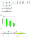
Figure 1
Figure 1
GPR110 transcripts and isoform 1 polypeptide. (A) Schematic of the four human GPR110 transcript isoforms, as gleaned from the UCSC genome web browser (chromosome 6, March 2006 version of the hg18 assembly). Isoform 1 encodes the full-length protein containing 14 exons. Isoform 2 contains an alternative splicing of exon 6 to exon 7a. Locations of in frame stop codons are shown for isoforms 3 and 4. (B) Relative expression of GPR110 isoforms 1-3 in human cell line PC-3. Transcript levels of isoforms 1 through 3 were measured by SYBR qPCR. Expression values were calculated relative to isoform 3 expression. Although qPCR analysis of the three GPR110 isoforms was done on the three prostate cells lines mentioned, as well as the three lung cell lines used in this study (A549, H460 and H23), we detected Isoform 2 only in PC-3 and LNCaP; and Isoform 3 only in PC-3. Thus, the data for all 3 isoforms are shown only for PC-3. (C) Schematic representation of the primary polypeptide structure of GPR110. SEA, SEA domain; GSIVA, amino acid sequence with potential for cleavage; GPS, GPS domain, with predicted cleavage at amino acid sequence HLT; 7TM, seven-span transmembrane domain. Blue triangles, predicted cleavage sites in the SEA and GPS domains; red triangles, locations of HA tags in isoform 1. Scale indicates peptide size, in kiloDaltons (kD).
Figure 2
Figure 2
Production of three GPR110 isoforms in human cell lines. (A) HA-tagged isoforms 1-3 were transiently expressed in HEK (lane1), HeLa (lane 2), and prostate line PC-3 (lane 3). Protein production detected by immunoblot with HA.11 antibody, which is specific to the HA tag; protein ladder molecular weights at the far left, in kD. Arrows point to the two major 80 and 100 kD molecular weight bands of isoform 1; and to a 30 kD band, representing a putative cleaved product. (B) isoforms 2 and 3, and (C) the four HA tagged isoform 1 proteins in HEK lysates (L) treated with (+) or without (-) glycosidase PNGaseF. Cterm, C-terminal HA-tagged isoform 1. Arrows point to the two major 60 and 75 kD bands after glycosidase treatment; and to a putative SEA cleavage product (in dotted box).
Figure 3
Figure 3
Isoform 1 is on the cell surface. Western blots of fractions of surface biotinylated HEK cells transfected with HA tagged GPR110 isoform 1 constructs (HA466 and HA1393), or with no tag (Iso1). (A) Developed with antibody to GAPDH (first blot), to integrin (second blot), or HA.11 antibody (third blot). L, lysate; U, unbound, and B, bound cell surface fraction. GAPDH is a cytosolic protein (arrow on the first blot); and integrin β1 is a surface protein (arrow on the second blot). Arrow on the third blot, 100 kD band of isoform 1. (B) Treatment of tagged GPR110 isoform 1 surface fractions with (+) or without (-) PNGaseF; developed with HA.11 antibody. Arrows point to the 60 and 75 kD bands, a result of the PNGaseF treatment; and to a 25 kD band, of the putative SEA fragment.
Figure 4
Figure 4
Isoforms 2 and 3 are on the cell surface. Western blots of fractions of surface biotinylated HEK cells transfected with C-terminal HA tagged isoforms 2 (Iso2) and 3 (Iso3), developed with antibody to GAPDH (first blot; arrow points to GAPDH), to integrin β1 (second blot; arrow points to integrin β1), and HA.11 antibody (third blot), respectively. pcDNA3.1, empty vector; L, lysate; U, unbound, and B, bound cell surface fraction.
Figure 5
Figure 5
Isoform 2 is secreted. Western blots of immunoprecipitates of cell culture media from GPR110 transfections; developed with antibody HA.11. (A) L, lysate; M, immunoprecipitate from media. HA466 and HA1393, two versions of HA-tagged isoform 1; Iso2-HA and Iso3-HA, HA-tagged isoform 2 and 3, respectively. Arrow points to the secreted form of isoform 2. (B) PNGaseF treatment of lysate and media of cells transfected with Iso2-HA. Arrow points to the deglycosylated isoform 2.
Figure 6
Figure 6
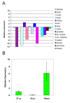
GPR110 is mainly expressed in the kidney. (A) Quantitative PCR measurements of expression across a human normal tissue panel, relative to the median gene expression of ~200 selected genes within each tissue. (B) Prostate samples from two young donors (24 and 27 yr) compared to the panel sample of RNA from the prostates of three 78-79 yr old men. The error bars represent technical replicates.
Figure 7
Figure 7
Gpr110 is an oncogene. Quantitative PCR measurements of Gpr110 expression in mouse T lymphomas generated by murine leukemia virus. Y-axis, relative expression on a logarithmic scale; x-axis, various T lymphomas. Tumors 754S and 3271S show overexpression of Gpr110. Tumor controls are RNAs from mouse tumors with no retroviral tags recovered from the Gpr110 locus. 'Normal' bar represents RNA from normal mouse spleen.
Figure 8
Figure 8
GPR110 mRNA overexpression in human lung cancer. GPR110 expression, as measured by qPCR with an exon junction 2-3 Taqman probe, and GUSB, glucuronidase beta, as a standard; in (A) lung cell lines A549, H23 and H460; y-axis, fluorescence; x-axis, number of cycles of amplification needed to reach a set value; in one set of (B) lung adenocarcinoma tumor samples and normal lung samples (Asterand); and in another set of (C) lung tumor and normal lung samples (Origene). The samples in (B) and (C) are separate cohorts, with (B) all representing adenocarcinomas. Tumors in (C) were sorted according to tumor type. The category of "other" includes adenosquamous, large cell, non-small, and sarcomatoid carcinomas. Numbers on the x-axis in (C) indicate well location on the Origene Lung TissueScan plate. Y-axis in (B) and (C) displays relative expression (2-ΔΔCt) normalized to average expression in the normal lung samples.
Figure 9
Figure 9
GPR110 protein overexpression in paraffin sections of lung cancer tissue. Immunohistochemistry on human lung cancer tissue arrays with consecutive sections, including normal controls, with antibodies LS-A2021 and LS-A2019. (A) Three peptides spanning amino acids 162-199 of GPR110. (B) Binding of LS-A2021 antibody to lung samples was competed with the three peptides. Peptide 2 decreased staining in GPR110-positive tissue samples, whereas lack of peptide, and peptides 1 and 3 did not. (C) Examples of cores staining with LS-A2021 only (type 1), with both antibodies (type 2), and with LS-A2019 only (type 3); there was no staining with IgG controls (not shown).
Figure 10
Figure 10
GPR110 overexpression in prostate benign hyperplasia and cancer. (A) GPR110 expression, in prostate cancer lines PC-3, LNCaP, and DU145, as measured by qPCR with an exon junction 2-3 Taqman probe; and GUSB, glucuronidase beta, as a standard. (B) Immunohistochemistry on two benign prostate hyperplasia cores, BPH-1 (upper) and BPH-2 (lower). Left, stained with antibody LS-A2021; right, consecutive sections of the same cores, stained with antibody to PSA. No staining was observed with secondary and irrelevant primary antibody (not shown). Note that the LS-A2021 positive cells are PSA negative, and vice versa (arrows). (C) Consecutive paraffin sections of two different prostate adenocarcinoma cores (1 and 2, left and right, respectively), stained with LS-A2021 antibody (upper two pictures) or LS-A2019 antibody (lower two pictures). Note that staining by the one antibody excludes staining by the other antibody.

References

    1. Filmore D. It's a GPCR world. Modern Drug Discovery. 2004;7(11):24–28.
    1. Levitin F, Stern O, Weiss M, Gil-Henn C, Ziv R, Prokocimer Z, Smorodinsky NI, Rubinstein DB, Wreschner DH. The MUC1 SEA module is a self-cleaving domain. J Biol Chem. 2005;280(39):33374–33386. doi: 10.1074/jbc.M506047200. - DOI - PubMed
    1. Khatri IA, Wang R, Forstner JF. SEA (sea-urchin sperm protein, enterokinase and agrin)-module cleavage, association of fragments and membrane targeting of rat intestinal mucin Muc3. Biochem J. 2003;372(Pt 1):263–270. doi: 10.1042/BJ20021333. - DOI - PMC - PubMed
    1. Krasnoperov VG, Bittner MA, Beavis R, Kuang Y, Salnikow KV, Chepurny OG, Little AR, Plotnikov AN, Wu D, Holz RW. alpha-Latrotoxin stimulates exocytosis by the interaction with a neuronal G-protein-coupled receptor. Neuron. 1997;18(6):925–937. doi: 10.1016/S0896-6273(00)80332-3. - DOI - PubMed
    1. Mikkers H, Allen J, Knipscheer P, Romeijn L, Hart A, Vink E, Berns A. High-throughput retroviral tagging to identify components of specific signaling pathways in cancer. Nat Genet. 2002;32(1):153–159. doi: 10.1038/ng950. - DOI - PubMed

Publication types

MeSH terms