. 2010;9(1):9.
doi: 10.1186/jbiol216. Epub 2010 Feb 11.

Madm (Mlf1 adapter molecule) cooperates with Bunched A to promote growth in Drosophila

Affiliations

Madm (Mlf1 adapter molecule) cooperates with Bunched A to promote growth in Drosophila

Silvia Gluderer et al. J Biol. 2010.

Abstract

Background: The TSC-22 domain family (TSC22DF) consists of putative transcription factors harboring a DNA-binding TSC-box and an adjacent leucine zipper at their carboxyl termini. Both short and long TSC22DF isoforms are conserved from flies to humans. Whereas the short isoforms include the tumor suppressor TSC-22 (Transforming growth factor-beta1 stimulated clone-22), the long isoforms are largely uncharacterized. In Drosophila, the long isoform Bunched A (BunA) acts as a growth promoter, but how BunA controls growth has remained obscure.

Results: In order to test for functional conservation among TSC22DF members, we expressed the human TSC22DF proteins in the fly and found that all long isoforms can replace BunA function. Furthermore, we combined a proteomics-based approach with a genetic screen to identify proteins that interact with BunA. Madm (Mlf1 adapter molecule) physically associates with BunA via a conserved motif that is only contained in long TSC22DF proteins. Moreover, Drosophila Madm acts as a growth-promoting gene that displays growth phenotypes strikingly similar to bunA phenotypes. When overexpressed, Madm and BunA synergize to increase organ growth.

Conclusions: The growth-promoting potential of long TSC22DF proteins is evolutionarily conserved. Furthermore, we provide biochemical and genetic evidence for a growth-regulating complex involving the long TSC22DF protein BunA and the adapter molecule Madm.


Figures
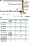
Figure 1
Figure 1
Long human TSC22DF isoforms can replace BunA function in Drosophila. (a) Schematic drawing of human and Drosophila TSC22DF proteins that were tested for their ability to rescue the lethality of bun mutants. The long isoforms possess two short conserved stretches named motif 1 and motif 2. Whereas BunA represents the long TSC22DF isoforms in Drosophila, BunB and BunC are two of the short isoforms. (b) Expression of long TSC22DF isoforms restores the viability of bun mutants. The quality of the rescue is indicated as a percentage of the expected Mendelian ratio. The Gal4 driver lines are ordered according to the strength of ubiquitous expression they direct during development, with arm-Gal4 being the weakest and Act5C-Gal4 the strongest driver line. In each experimental cross, n ≥ 200 progeny flies were analyzed. Leaky expression, without Gal4; 1 c and 2 c, one or two copies of the respective UAS construct. The ZH-attP-86Fb integration site seems to mediate strong expression as the UAS-attB-bunA constructs (ORF and cDNA) do not need to be driven by a Gal4 line for rescue, in contrast to the UAS-bunA construct (cDNA) generated by standard P-element-mediated germline transformation (inserted non-site-specifically on chromosome III). Note that too high expression of long TSC22DF members is harmful to flies. In a wild-type background, Act5C-Gal4-directed expression (n ≥ 200) of TSC22D2 and of bunA ORF kills the animals (0% survival). Expression from the bunA cDNA construct produces few escapers (3%), whereas expression from the bunA cDNA P-element construct and of TSC22D4 results in semi-viability (14% and 69%, respectively). Only TSC22D1.1 can be expressed by Act5C-Gal4 without compromising survival (>80%). Thus, there appears to be an optimal range of long TSC22DF concentration for viability.
Figure 2
Figure 2
Madm interacts biochemically with BunA. (a) Western blot showing that endogenous BunA is pulled down together with HA-Madm. Anti-HA beads were used to capture either HA-Madm or HA-eGFP as a negative control, respectively. A tenth of the cell lysate was used for the input control. (b, c) Co-localization studies of BunA and Madm in Drosophila S2 cells. In (b-b") a stable cell line capable of producing GFP-BunA in every cell was transiently transfected with a plasmid leading to expression of HA-Madm in a subset of cells (and vice versa in c-c"). Co-overexpression of GFP-BunA influences the localization of HA-Madm, resulting in a less dispersed pattern (c-c"). (d) GFP-BunA co-localizes with the Golgi marker GMAP210 (Golgi microtubule-associated protein of 210 kDa) [38]. (e, f) Schematic drawing of BunA (e) and Madm (f) constructs tested in Y2H and co-IP assays for an interaction with full-length Madm and BunA, respectively. The results of the Y2H and co-IP experiments are summarized on the left [see Additional files 2 and 3 for the primary results]. The physical interaction of BunA and Madm is mediated by a short protein sequence encompassing the conserved motif 2 in BunA and a carboxy-terminal sequence in Madm, respectively [see Additional file 4 for alignments].
Figure 3
Figure 3
A genetic eyFLP/FRT-based screen in Drosophila identifies Madm as a positive growth regulator. (a-d) Dorsal view of mosaic heads generated by means of the eyFLP/FRT system. (a) The isogenized FRT82 chromosome used in the genetic screen produces a control mosaic head. (b, c) Heads largely homozygous mutant for an EMS-induced Madm mutation display a pinhead phenotype that can be reverted by one copy of a genomic Madm rescue construct (d). (e) Graphic representation of the Drosophila Madm protein (top) and gene (bottom). In the protein, the BunA-binding region and the NES and NLS sequences are indicated (netNES 1.1 [63], ELM [64], PredictNLS [65]). The seven alleles isolated in the genetic screen and the sites of their EMS-induced mutations are in red. Amino acid changes in the protein are indicated. In alleles 3Y2 and 7L2, the first nucleotide downstream of the first Madm exon is mutated, thus disrupting the splice donor site. In allele 2D2, a deletion causes a frameshift after amino acid 385, resulting in a premature translational stop after an additional 34 amino acids. Alleles 3Y2, 4S3, and 7L2 lead to a pinhead phenotype of intermediate strength (b) whereas 2D2, 2U3, and 3G5 produce a stronger pinhead phenotype (c). The hypomorphic allele 3T4 generates a weak pinhead phenotype (data not shown). Genotypes of the flies shown are: (a) y, w, eyFlp/y, w; FRT82B/FRT82B, w+, cl3R3; (b, c) y, w, eyFlp/y, w; FRT82B, Madm7L2 or 3G5/FRT82B, w+, cl3R3; (d) y, w, eyFlp/y, w; gen.Madm(LCQ139)/+; FRT82B, Madm3G5/FRT82B, w+, cl3R3.
Figure 4
Figure 4
The Madm loss- or reduction-of-function phenotypes strongly resemble bunA phenotypes. (a-c) Scanning electron micrographs of eyFLP/FRT mosaic eyes. (d) Madm mosaic heads (b, c) contain significantly fewer ommatidia than control mosaic heads (a) (n = 6). (a'-c') Images of tangential eye sections showing that Madm mutant (unpigmented) ommatidia (b', c') display an autonomous reduction in rhabdomere size relative to wild-type sized (pigmented) ommatidia. Furthermore, differentiation defects such as misrotation and missing photoreceptors are observed in Madm mutant ommatidia. Clones were induced 24-48 h after egg deposition using the hsFLP/FRT technique. (e) Rhabdomere size of Madm-mutant ommatidia is significantly reduced (by 29-56%). The area enclosed by rhabdomeres of photoreceptors R1-R6 in unpigmented mutant ommatidia was compared to the area measured in pigmented wild-type sized ommatidia. For each genotype, three pairs of ommatidia without differentiation defects from three different eye sections were measured (n = 9). Significant changes are marked by asterisks, **p < 0.01 and ***p < 0.001 (Student's t-test) in (d) and (e). (f) Heteroallelic combinations of the hypomorphic Madm allele 3T4 produce few viable small flies (<10% of the expected Mendelian ratio) that can be rescued by one copy of a genomic Madm rescue construct. (g) The dry weight of Madm hypomorphic females is reduced by 37% compared to control flies (Df/+). One copy of a genomic rescue construct restores normal weight. The genomic rescue construct has no significant dominant effect on dry weight ('rescue Df/+' females do not significantly differ from 'Df/+' females). n = 15, except for Df/3T4 (n = 9). (h) Tangential section of an eye from a Madm hypomorphic mutant female displaying rotation defects (yellow asterisk), missing rhabdomeres (green asterisk), and cell-fate transformations (red asterisk). (i) Wings of hypomorphic Madm males exhibiting wing notches and an incomplete wing vein V (arrows). Genotypes are: (a, a') y, w, eyFlp or hsFlp/y, w; FRT82B/FRT82B, w+, cl3R3 or M. (b, b', c, c') y, w, eyFlp or hsFlp/y, w; FRT82B, Madm7L2 or 3G5/FRT82B, w+, cl3R3 or M; (Df/+) y, w; FRT82B/Df(3R)Exel7283; (Df/3T4) y, w; FRT82B, Madm3T4/Df(3R)Exel7283; (rescue Df/3T4) y, w; gen.Madm(LCQ139)/+; FRT82B, Madm3T4/Df(3R)Exel7283; (rescue Df/+) y, w; gen.Madm(LCQ139)/+; FRT82B/Df(3R)Exel7283.
Figure 5
Figure 5
Co-overexpression of Madm and bunA causes overgrowth. (a-d) Scanning electron micrographs of adult eyes as a readout for the consequences of overexpression of bunA and Madm under the control of the GMR-Gal4 driver line late during eye development. Whereas expression of (b) bunA or (c) Madm singly does not cause a size alteration compared to the control (a), overexpression of both leads to increased eye size (d). (e) The size increase on bunA and Madm coexpression is due to larger ommatidia (Student's t-test, n = 9, ***p < 0.001). (f-i) The growth-promoting effect of bunA and Madm co-overexpression is also observed in the wing. Single expression of either (g, g') bunA or (h, h') Madm during wing development (by means of C10-Gal4) does not change wing size or curvature visibly. However, their combined expression causes a slight overgrowth of the tissue between the wing veins, resulting in a wavy wing surface and wing bending (i'), manifested as folds between wing veins in (i) (arrows). Genotypes are: (a) y, w; GMR-Gal4/UAS-eGFP; UAS-lacZ/+; (b) y, w; GMR-Gal4/UAS-eGFP; UAS-bunA/+; (c) y, w; GMR-Gal4/UAS-Madm; UAS-lacZ/+; (d) y, w; GMR-Gal4/UAS-Madm; UAS-bunA/+; (f) y, w; UAS-eGFP/+; C10-Gal4/UAS-lacZ; (g) y, w; UAS-eGFP/+; C10-Gal4/UAS-bunA; (h) y, w; UAS-Madm/+; C10-Gal4/UAS-lacZ; (i) y, w; UAS-Madm/+; C10-Gal4/UAS-bunA.

Similar articles

Cited by

References

    1. Shibanuma M, Kuroki T, Nose K. Isolation of a gene encoding a putative leucine zipper structure that is induced by transforming growth factor beta 1 and other growth factors. J Biol Chem. 1992;267:10219–10224. - PubMed
    1. Iida M, Anna CH, Holliday WM, Collins JB, Cunningham ML, Sills RC, Devereux TR. Unique patterns of gene expression changes in liver after treatment of mice for 2 weeks with different known carcinogens and non-carcinogens. Carcinogenesis. 2005;26:689–699. doi: 10.1093/carcin/bgi005. - DOI - PubMed
    1. Nakashiro K, Kawamata H, Hino S, Uchida D, Miwa Y, Hamano H, Omotehara F, Yoshida H, Sato M. Down-regulation of TSC-22 (transforming growth factor beta-stimulated clone 22) markedly enhances the growth of a human salivary gland cancer cell line in vitro and in vivo. Cancer Res. 1998;58:549–555. - PubMed
    1. Rentsch CA, Cecchini MG, Schwaninger R, Germann M, Markwalder R, Heller M, Pluijm G van der, Thalmann GN, Wetterwald A. Differential expression of TGFbeta-stimulated clone 22 in normal prostate and prostate cancer. Int J Cancer. 2006;118:899–906. doi: 10.1002/ijc.21449. - DOI - PubMed
    1. Shostak KO, Dmitrenko VV, Vudmaska MI, Naidenov VG, Beletskii AV, Malisheva TA, Semenova VM, Zozulya YP, Demotes-Mainard J, Kavsan VM. Patterns of expression of TSC-22 protein in astrocytic gliomas. Exp Oncol. 2005;27:314–318. - PubMed

Publication types

MeSH terms