. 2010 May 15;51(1):53-62.
doi: 10.1016/j.neuroimage.2010.02.006. Epub 2010 Feb 10.

The interrelationship of dopamine D2-like receptor availability in striatal and extrastriatal brain regions in healthy humans: a principal component analysis of [18F]fallypride binding

Affiliations

The interrelationship of dopamine D2-like receptor availability in striatal and extrastriatal brain regions in healthy humans: a principal component analysis of [18F]fallypride binding

David H Zald et al. Neuroimage. .

Abstract

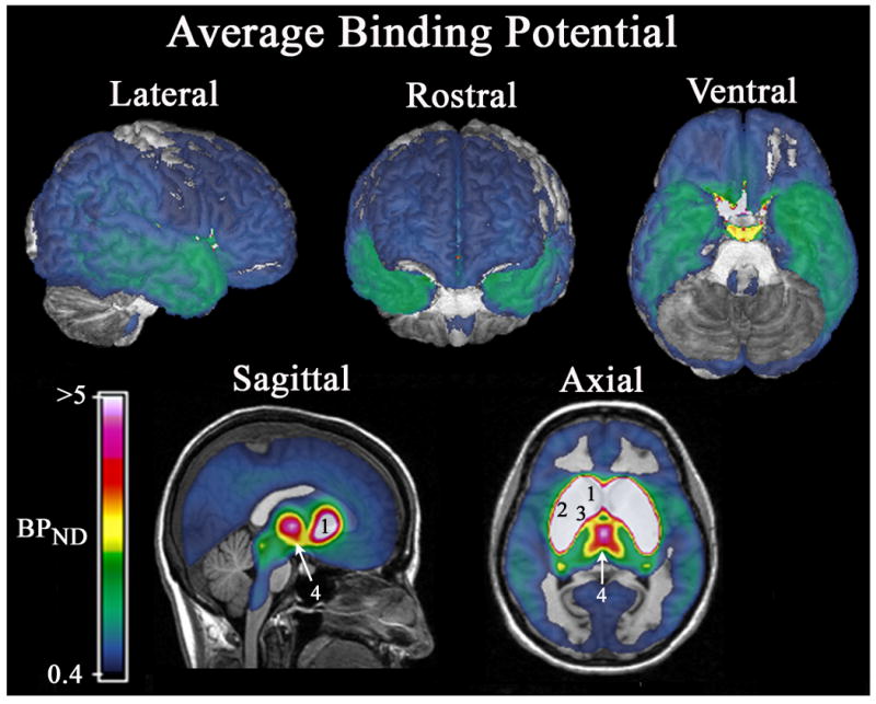
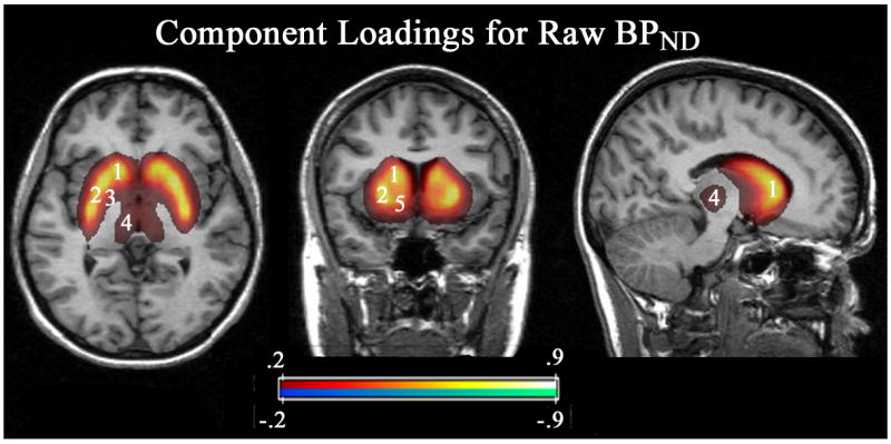
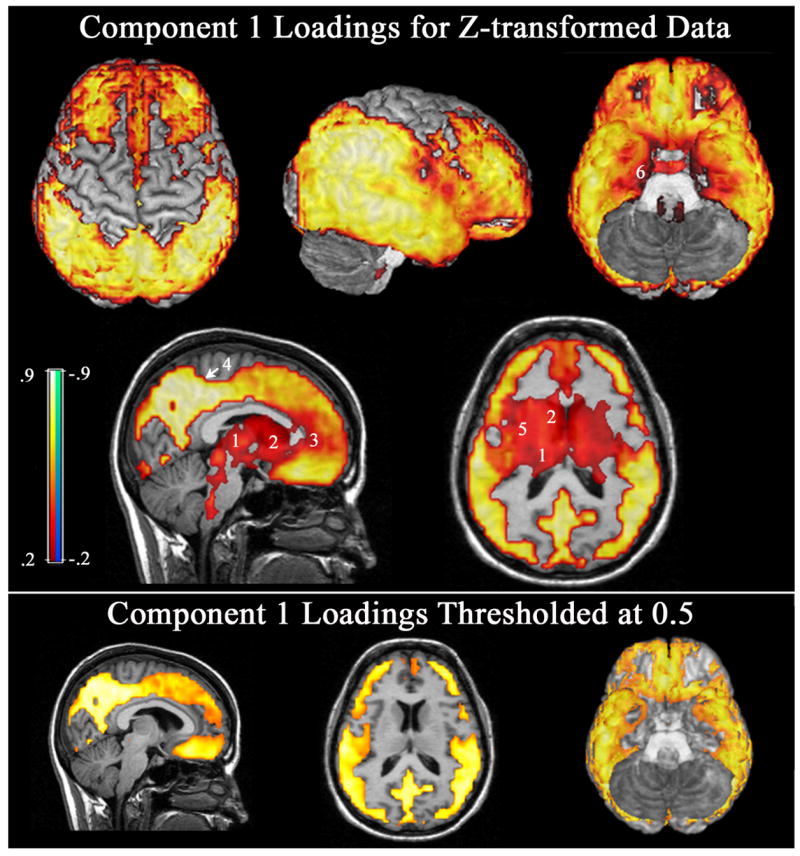
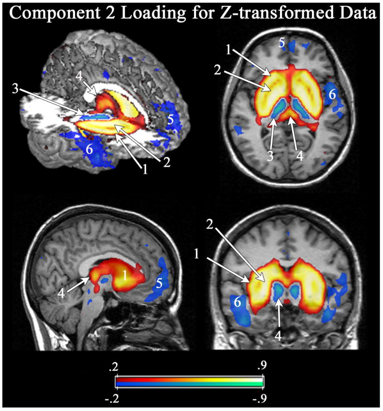
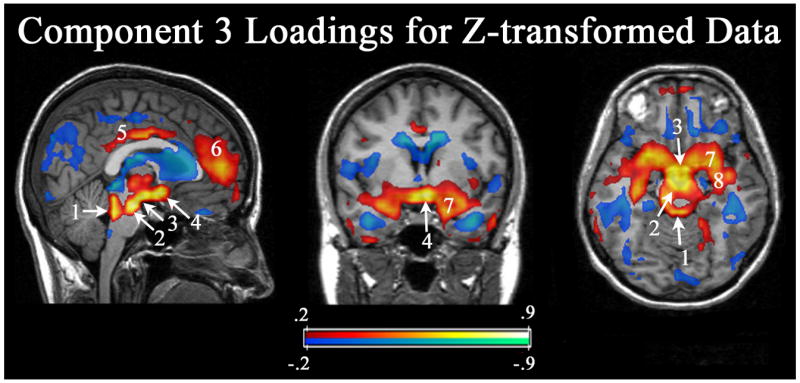
Individual differences in dopamine D2-like receptor availability arise across all brain regions expressing D2-like receptors. However, the interrelationships in receptor availability across brain regions are poorly understood. To address this issue, we examined the relationship between D2-like binding potential (BPND) across striatal and extrastriatal regions in a sample of healthy participants. PET imaging was performed with the high affinity D2/D3 ligand [18F]fallypride in 45 participants. BPND images were submitted to voxel-wise principal component analysis to determine the pattern of associations across brain regions. Individual differences in D2-like BPND were explained by three distinguishable components. A single component explained almost all of the variance within the striatum, indicating that individual differences in receptor availability vary in a homogenous manner across the caudate, putamen, and ventral striatum. Cortical BPND was only modestly related to striatal BPND and mostly loaded on a distinct component. After controlling for the general level of cortical D2-like BPND, an inverse relationship emerged between receptor availability in the striatum and the ventral temporal and ventromedial frontal cortices, suggesting possible cross-regulation of D2-like receptors in these regions. The analysis additionally revealed evidence of: (1) a distinct component involving the midbrain and limbic areas; (2) a dissociation between BPND in the medial and lateral temporal regions; and (3) a dissociation between BPND in the medial/midline and lateral thalamus. In summary, individual differences in D2-like receptor availability reflect several distinct patterns. This conclusion has significant implications for neuropsychiatric models that posit global or regionally specific relationships between dopaminergic tone and behavior.


Figures

Figure 1
Figure 1
Average [18F]Fallypride BPND. The data represents the average of all 45 subjects who participated in the present study. The data were thresholded to only show voxels with an average BPND ≥ 0.4. The BPND maps were overlaid on cortical surface renderings (top row) or sagittal and axial slices (bottom row) of a template brain. Note, the selected color scale emphasizes regional differences in BPND within cortical areas at the expense of viewing differences in the basal ganglia regions. Because the caudate, putamen, ventral striatum and globus pallidus all possess BPND > 5, it is not possible to distinguish their boundaries with this color scale. In addition to cortical areas, the sagittal slice in the second row shows a band of binding along the midline of the dorsal aspect of the brainstem which includes the colliculi, the raphe nucleus, and the nucleus of the solitary tract, all of which have previously been identified to possess D2 receptors in autoradiographic studies (Yokoyama et al., 1994; Hurd et al., 2001). Number labels: 1) caudate, 2) putamen, 3) globus pallidus, 4) thalamus.
Figure 2
Figure 2
Component loadings for the single Component that arose from the raw BPND data. Strongly positive loadings emerged throughout the striatum, with highest loadings in the dorsal caudate and putamen, and slightly lower loadings in the ventral striatum. Outside of the striatum, only the globus pallidus and thalamus also loaded on this component, but at much weaker levels than the striatum. The sagittal slice runs through the caudate and also displays part of the thalamus. In the present figure and all subsequent figures, the data were thresholded to only show component loadings ≥ .2 or ≤ -.2. Number labels: 1) caudate, 2) putamen, 3) globus pallidus, 4) thalamus, 5) ventral striatum
Figure 3
Figure 3
Principal Component #1 from the z-transformed data. The 1st component is marked by a broad pattern of positive associations throughout widespread areas of cortex. For purpose of display voxel-wise loadings are overlaid on surface renderings of the dorsal, lateral and ventral surface of the brain (left to right top row), as well as a midline sagittal and an axial slice at the level of the dorsal striatum (2nd row). The strongest loadings localized to the posterior cingulate/precuneus region, with additional strong loadings in parietal cortex, anterior and ventral temporal cortex, and portions of the frontal lobe. The bottom of the figure shows the sagittal and axial slices and the ventral surface rendering after thresholding to only show loadings ≥ .5 in order to more clearly delineate areas with strong loadings vs. areas with more moderate loadings. Although positive loadings were broad, several areas show only modest or moderate loadings on the 1st component, including including the pregenual cingulate region and infero-medial temporal cortex (which drop out in the thresholded images). Moderate positive loadings can also be seen in the thalamus, and striatum, which also drop out when thresholding is applied. Similarly, the sagittal slice in the 2nd row shows moderate loadings in the dorsal aspect of the brainstem (posterior pons), where binding occurred in the vicinity of the raphe nuclei, nucleus of the solitary tract, and locus coeruleus, but did not surpass the 0.5 threshold. Number labels: 1) thalamus, 2) caudate, 3) pregenual cingulate, 4) precuneus, 5) putamen, 6) medial temporal cortex.
Figure 4
Figure 4
Principal Component #2. The 2nd component involves strong positive loadings in the basal ganglia and moderate loadings in the posterior-medial thalamus, and SN/VTA, but negative loadings in the anterior-lateral thalamus and cortical regions. Number labels: 1) striatum, 2) globus pallidum, 3) anterior-lateral thalamus, 4) posterior-medial thalamus, 5) ventromedial PFC, 6) lateral temporal cortex, and 7) substantia nigra/ventral tegmental area.
Figure 5
Figure 5
Principal Component #3. The 3rd component produced its highest loadings in the DA midbrain, the mammillary bodies and the hypothalamus. Note in the coronal and axial slices, a large area of the basal forebrain, including the extended amygdala also positively loads on this component. The axial slice passes through the mammillary bodies, which is slightly superior to the peak loadings in the substantia nigra/ventral tegmental area. Number labels: 1) colliculus/raphe, 2) substantia nigra/ventral tegmental area, 3) mammillary bodies, 4) hypothalamus, 5) posterior/mid cingulate, 6) medial frontal cortex, 7) amygdala, and 8) hippocampus.

Similar articles

Cited by

References

    1. Abi-Dargham A, Mawlawi O, Lombardo I, Gil R, Martinez D, Huang YY, Hwang DR, Keilp J, Kochan L, Van Heertum R, Gorman JM, Laruelle M. Prefrontal dopamine D-1 receptors and working memory in schizophrenia. Journal of Neuroscience. 2002;22:3708–3719. - PMC - PubMed
    1. Bannon MJ, Roth RH. Pharmacology of Mesocortical Dopamine Neurons. Pharmacological Reviews. 1983;35:53–68. - PubMed
    1. Buchsbaum MS, Christian BT, Lehrer DS, Narayanan TK, Shi B, Mantil J, Kemether E, Oakes TR, Mukherjee J. D2/D3 dopamine receptor binding with [F-18]fallypride in thalamus and cortex of patients with schizophrenia. Schizophrenia Research. 2006;85:232–244. - PubMed
    1. Bull DR, Bakhtiar R, Sheehan MJ. Characterization of dopamine autoreceptors in the amygdala: a fast cyclic voltammetric study in vitro. Neuroscience Letters. 1991;134:41–44. - PubMed
    1. Clatworthy PL, Lewis SJ, Brichard L, Hong YT, Izquierdo D, Clark L, Cools R, Aigbirhio FI, Baron JC, Fryer TD, Robbins TW. Dopamine release in dissociable striatal subregions predicts the different effects of oral methylphenidate on reversal learning and spatial working memory. J Neurosci. 2009;29:4690–4696. - PMC - PubMed

Publication types

Substances