

doi: 10.1101/pdb.prot5384.
DNase-seq: a high-resolution technique for mapping active gene regulatory elements across the genome from mammalian cells
Affiliations
- PMID: 20150147
- PMCID: PMC3627383
- DOI: 10.1101/pdb.prot5384
Item in Clipboard
DNase-seq: a high-resolution technique for mapping active gene regulatory elements across the genome from mammalian cells
Cold Spring Harb Protoc.
2010 Feb.
No abstract available
Figures

Flow chart of DNase-seq protocol. Briefly, cells are lysed with detergent to release nuclei, and the nuclei are digested with optimal concentrations of DNase I. DNase I digested DNA is embedded in low-melt gel agarose plugs to reduce additional random shearing. DNA (while still in the plugs) are then blunt-ended, extracted and ligated to biotinylated linker 1 (represented by red bars in the figure). Excess linker is removed by gel purification, and biotinylated fragments (Linker 1 plus 20 bases of genomic DNA) are digested with MmeI, and captured by streptavidin-coated Dynal beads (represented by brown balls). Linker 2 (represented by the blue bars) is ligated to the 2 base overhang generated by MmeI, and the ditagged 20 bp DNAs are amplified by PCR and sequenced by Illumina/Solexa.

Pulsed field gel picture of DNaseI digested DNA isolated from Human Umbilical Vein Endothelial cells (HUVEC). Ideal PFG displays gradual and consistent changes in high molecular weight DNA fragment sizes as DNase I concentrations increase.
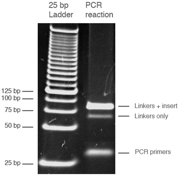
Gel picture of PCR reactions. 1 μL of 25 bp ladder and 30 μL of PCR reaction were loaded on a 4–20% TBE-PAGE gel. The linkers with insert band is 86 bp, the linker-only band is 66 bp, and the PCR primers are 20–30 bases.
References
-
- Gross DS, Garrard WT. Nuclease hypersensitive sites in chromatin. Annu Rev Biochem. 1988;57:159–97. - PubMed
Publication types
MeSH terms
Substances
Grants and funding
LinkOut - more resources
Full Text Sources
Other Literature Sources