. 2010 May 1;19(9):1791-804.
doi: 10.1093/hmg/ddq060. Epub 2010 Feb 11.

AP-2alpha knockout mice exhibit optic cup patterning defects and failure of optic stalk morphogenesis

Affiliations

AP-2alpha knockout mice exhibit optic cup patterning defects and failure of optic stalk morphogenesis

Erin A Bassett et al. Hum Mol Genet. .

Abstract

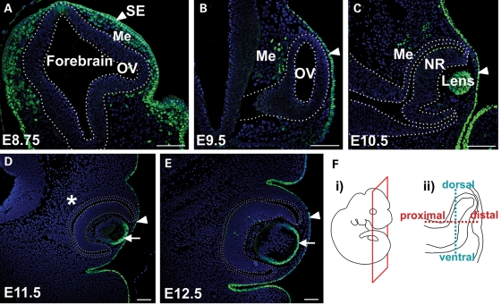
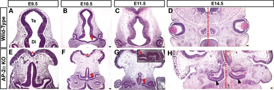
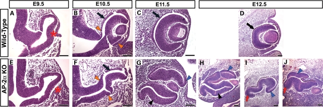
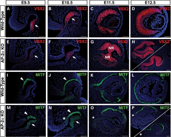
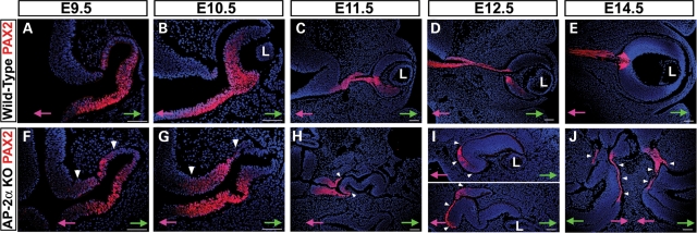
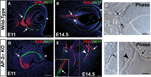
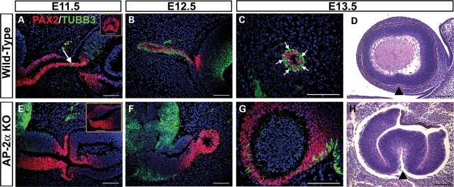
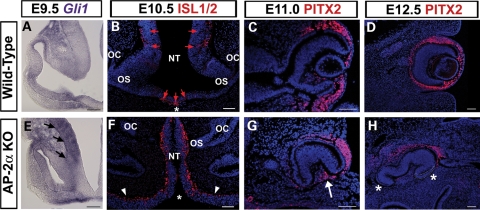
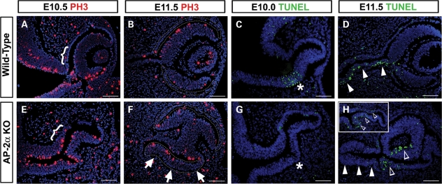
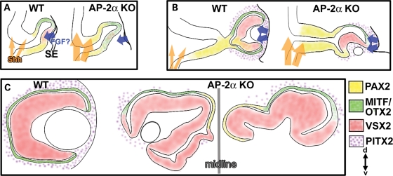
Appropriate development of the retina and optic nerve requires that the forebrain-derived optic neuroepithelium undergoes a precisely coordinated sequence of patterning and morphogenetic events, processes which are highly influenced by signals from adjacent tissues. Our previous work has suggested that transcription factor activating protein-2 alpha (AP-2alpha; Tcfap2a) has a non-cell autonomous role in optic cup (OC) development; however, it remained unclear how OC abnormalities in AP-2alpha knockout (KO) mice arise at the morphological and molecular level. In this study, we show that patterning and morphogenetic defects in the AP-2alpha KO optic neuroepithelium begin at the optic vesicle stage. During subsequent OC formation, ectopic neural retina and optic stalk-like tissue replaced regions of retinal pigment epithelium. AP-2alpha KO eyes also displayed coloboma in the ventral retina, and a rare phenotype in which the optic stalk completely failed to extend, causing the OCs to be drawn inward to the midline. We detected evidence of increased sonic hedgehog signaling in the AP-2alpha KO forebrain neuroepithelium, which likely contributed to multiple aspects of the ocular phenotype, including expansion of PAX2-positive optic stalk-like tissue into the OC. Our data suggest that loss of AP-2alpha in multiple tissues in the craniofacial region leads to severe OC and optic stalk abnormalities by disturbing the tissue-tissue interactions required for ocular development. In view of recent data showing that mutations in human TFAP2A result in similar eye defects, the current findings demonstrate that AP-2alpha KO mice provide a valuable model for human ocular disease.


Figures

Figure 1.
Figure 1.
Expression pattern of AP-2α protein during early eye development. Immunofluorescence using an anti-AP-2α antibody (green) on sections of wild-type embryos counterstained with DAPI (blue). (AE) Coronal paraffin sections of mouse embryo heads at the developmental stages indicated. Outlined region in A indicates the forebrain neuroepithelium, including an evaginating OV. Outlined regions in B–E demarcate the OV/OC. Arrowheads in A–E denote surface ectoderm, whereas arrows in D, E denote anterior lens epithelium. AP-2α was not detected in the periocular mesenchyme beyond E10.5 (asterisk in D). (F) Schematics showing the plane of section (i) used on all embryos throughout this study except for sagittal sections in Fig. 7, and the dorsal-ventral and proximal-distal axes in the developing optic neuroepithelium (ii). SE, surface ectoderm; OV, optic vesicle; Me, mesenchyme; NR, neural retina. Scalebars, 100 µm.
Figure 2.
Figure 2.
Defects in AP-2α KO cranial and ocular regions arise during early eye morphogenesis. (AH) Coronal H&E-stained paraffin sections of wild-type (A–D) and AP-2α KO (E–H) embryo heads at the stages indicated. Red asterisks (E, F) denote the open cranial neural tube in AP-2α KO mice. Red arrowheads (B, F, G) show the shortened, thickened optic stalk in AP-2α KOs compared with controls. Inset in G illustrates example of a severely turned AP-2α KO eye. By E14.5, the AP-2α KO eyes are adjacent to the midline (dotted red lines in D, H). Black arrowheads (H) show duplicated NR in the ventral AP-2α KO OC. Te, telencephalon; Di, diencephalon. Scalebars, 100 µm.
Figure 3.
Figure 3.
AP-2α KO eye defects begin at OV stage. (AJ) Coronal H&E-stained paraffin sections of wild-type (A–D) and AP-2α KO (E–J) embryo heads at the stages indicated. (A, E) Red asterisks denote the mispositioning of the AP-2α KO OV at E9.5, which was not oriented correctly with respect to the surface ectoderm. (B, F) Black arrows point to regions of the wild-type and AP-2α KO outer OC that are similar in thickness, whereas orange arrowheads point to regions of presumptive RPE adjacent to the optic stalk that are thickened in AP-2α KOs compared with controls. (C, D, G–J) Although the RPE formed a clear monolayer at E11.5 and onwards in wild-type eyes (black arrows in C, D), regions of the outer OC layer in AP-2α KOs were visibly thickened in both the dorsal OC (blue arrowheads in G–J) and ventral OC (black arrowheads in G, H). Red arrows in I, J show AP-2α KO eyes with an underdeveloped ventral retina. Scalebars, 100 µm.
Figure 4.
Figure 4.
Ocular patterning defects and formation of a duplicated NR in AP-2α KOs. Immunofluorescence using anti-VSX2/CHX10 (red) and anti-MITF (green) on coronal sections of wild-type (AD, IL) and AP-2α KO (EH, MP) embryo heads counterstained with DAPI (blue), at the stages indicated. Examples of two E12.5 eyes are shown (H, P), to illustrate the variability in the extent and location of ectopic NR tissue. (A, B, E, F) Arrows point to VSX2 expression in NR domain, which was reduced in AP-2α KOs compared with controls at E9.5 and E10.5. (I, J, M, N) Arrowheads denote MITF expression in the wild-type and AP-2α KO RPE domain, whereas arrows point to persistent MITF expression in the NR domain of AP-2α KOs at E9.5 and E10.5. (C, D, G, H) Outlined regions illustrate ectopic VSX2 expression in the AP-2α KO duplicated NR. (K, L, O, P) Outlined regions show absent or reduced MITF expression in thickened regions of the AP-2α KO outer OC. Asterisks (N–P) indicate additional regions of the outer OC adjacent to the midline that lack MITF expression. Scalebars, 100 µm.
Figure 5.
Figure 5.
PAX2 expansion into RPE domain in AP-2α KOs. Immunofluorescence using anti-PAX2 (red) on coronal sections of wild-type (AE) and AP-2α KO (FJ) embryo heads counterstained with DAPI (blue), at the stages indicated. (A, B, F, G) White arrowheads show dorsal expansion of PAX2 expression in AP-2α KO eyes compared with wild-type eyes at E9.5 and E10.5. (C–E, H–J) Although PAX2 became confined to the optic stalk and newly formed optic disk in wild-type embryos, white arrowheads denote expansion of the PAX2 expression domain into regions of presumptive RPE closest to the midline in AP-2α KOs. Refer to Fig. 2H for an H&E stain of the same embryo depicted in J. Pink and green arrows in A–J point towards midline and surface ectoderm, respectively. L, lens. Scalebars, 100 µm.
Figure 6.
Figure 6.
The PAX2-MITF border is shifted into the OC in AP-2α KOs. (A, B, D, E) Co-immunostaining of PAX2 (red) and MITF (green) on coronal sections of wild-type (A, B) and AP-2α KO (D, E) embryo heads counterstained with DAPI (blue), at the stages indicated. White arrows (A, B, D, E) denote the PAX2-MITF borders, whereas white arrowhead (E) indicates overlap of PAX2-positive and MITF-positive cells. Asterisks (A, B, D) mark the surface ectoderm. Dashed red line (E) marks the midline. Boxed area in E is magnified in inset. (C, F) Corresponding phase views of the eyes in B and E, respectively. Black arrows point to the border between pigmented versus non-pigmented cells, and black arrowhead denotes lack of pigmentation in area of PAX2-MITF overlap. Refer to Fig. 2H for an H&E stain of the same embryo depicted in E and F. Scalebars, 100 µm.
Figure 7.
Figure 7.
Optic nerve development and optic fissure closure are impaired in AP-2α KOs. (AC, EG) Co-immunostaining of PAX2 (red) and neuron specific class III β-tubulin (TUBB3; green) on coronal (A, B, E, F) and sagittal (C, G) sections of wild-type (A–C) and AP-2α KO (E–G) embryo heads counterstained with DAPI (blue), at the stages indicated. Arrow (A) points to TUBB3-positive RGC axons entering the PAX2-positive optic stalk and extending towards the diencephalon, which was not observed in AP-2α KOs (E, F). Insets (A, E) show view of PAX2-stained optic stalk from sagittal sections of wild-type (A) or AP-2α KO (E) embryos at E11.5. Arrows in C show examples of TUBB3-positive RGC axon bundles adjacent to PAX2-positive glial precursors in the wild-type E13.5 optic stalk. (D, H) Sagittal H&E-stained sections of AP-2α KO and control littermate embryo heads. By E13.5, the optic fissure margins in wild-type eyes had fully fused along the ventral OC, but were consistently open in AP-2α KOs (black arrowheads). Scalebars, 100 µm.
Figure 8.
Figure 8.
Altered gene expression in neural tube and POM of AP-2α KOs. (A, E) In situ hybridization of Gli1 (purple) on coronal sections of wild-type (A) and AP-2α KO (E) embryo heads at E9.5. Black arrows in E indicate increased signal in AP-2α KO neural tube and proximal OV. (BD, FH) Immunofluorescence using anti-ISL1/2 (B, F) and anti-PITX2 (C, D, G, H) (red) on coronal sections of wild-type (B–D) and AP-2α KO (F–H) embryo heads counterstained with DAPI (blue), at the stages indicated. Asterisks in B and F indicate the closed versus open neural tube in wild-type and AP-2α KO, respectively. Red arrows in B show ISL1-positive cells in wild-type forebrain. Arrowheads in F denote overgrown neuroepithelium in AP-2α KO. Arrow in G indicates PITX2-positive cells inside the AP-2α KO OC at E11. Asterisks in H denote regions adjacent to the mutant OC that contain either fewer or no cells expressing PITX2. Outlined area in H indicates ectopic NR. OS, optic stalk; OC, optic cup; NT, neural tube. Scalebars, 100 µm.
Figure 9.
Figure 9.
Changes in cell proliferation and apoptosis in AP-2α KO eyes. (A, B, E, F) Immunofluorescence using the mitotic marker PH3 (red) on coronal sections of wild-type (A, B) and AP-2α KO (E, F) embryo heads counterstained with DAPI (blue), at the stages indicated. Brackets (A, E) indicate the region at the optic stalk-RPE border in which AP-2α KOs show an increase in PH3-labeled cells compared with controls. Outlines (B, F) denote the presumptive RPE territory, and arrows in F point to a highly proliferative area in the AP-2α KO outer OC. (C, D, G, H) Apoptotic cells (green) detected using the TUNEL assay on coronal sections of wild-type (C, D) and AP-2α KO (G, H) embryo heads counterstained with DAPI (blue), at the stages indicated. Asterisks (C, G) indicate the previously reported ventral region of apoptosis (C), which was not detected in AP-2α KOs (G). Solid arrowheads (D, H) point to the E11.5 optic stalk, in which AP-2α KOs lacked the apoptotic cells seen in wild-type eyes. Inset in H shows example of a second AP-2α KO OC at E11.5. Open arrowheads (H) show examples of cells undergoing programmed death in the AP-2α KO OCs. Scalebars, 100 µm.
Figure 10.
Figure 10.
Summary of ocular neuroepithelium development in AP-2α germ-line KOs. All representative eyes depict coronal sections oriented according to the dorsal (d)–ventral (v) axis at bottom right. (A) At the OV stage, the forebrain neuroepithelium exhibits increased responsiveness to SHH, suggestive of increased activation of the SHH pathway (orange arrows). Deficient contact between the OV and surface ectoderm in AP-2α KOs disrupts lens formation and causes a persistence of RPE determinants (green) in the presumptive NR, likely due to the fact the distal OV is not close enough to NR-promoting signals from the surface ectoderm (possibly FGF; blue arrows). (B) The AP-2α KO OC typically develops in a position that is rotated to bring a portion of the dorsal presumptive RPE in apposition to the surface ectoderm. This rotation causes a segment of dorsal RPE to be transiently exposed to surface ectoderm-derived signals, which favors NR characteristics (red). As a result of failed optic stalk extension, the OC is pulled towards the midline, a source of SHH signals that presumably maintain PAX2 expression (yellow) in the proximal OC. The AP-2α KO eyes are also mispositioned with respect to PITX2-positive POM cells (stippled, purple), which further affects exposure to inductive signals. (C) The resulting AP-2α KO OCs sit adjacent to the midline and display variable defects, including lack of ventral retina tissue or a duplicated NR throughout the ventral RPE domain, regions of dorsal RPE-to-NR conversion and no optic stalk.

Similar articles

Cited by

References

    1. Wilson S.W., Houart C. Early steps in the development of the forebrain. Dev. Cell. 2004;6:167–181. - PMC - PubMed
    1. Rubenstein J.L., Beachy P.A. Patterning of the embryonic forebrain. Curr. Opin. Neurobiol. 1998;8:18–26. - PubMed
    1. Gage P.J., Rhoades W., Prucka S.K., Hjalt T. Fate maps of neural crest and mesoderm in the mammalian eye. Invest. Ophthalmol. Vis. Sci. 2005;46:4200–4208. - PubMed
    1. Chow R.L., Lang R.A. Early eye development in vertebrates. Annu. Rev. Cell. Dev. Biol. 2001;17:255–296. - PubMed
    1. Yang X.J. Roles of cell-extrinsic growth factors in vertebrate eye pattern formation and retinogenesis. Semin. Cell Dev. Biol. 2004;15:91–103. - PMC - PubMed

Publication types

MeSH terms