. 2010 May;298(5):G643-56.
doi: 10.1152/ajpgi.00260.2009. Epub 2010 Feb 11.

Interstitial cells of Cajal in the cynomolgus monkey rectoanal region and their relationship to sympathetic and nitrergic nerves

Affiliations

Interstitial cells of Cajal in the cynomolgus monkey rectoanal region and their relationship to sympathetic and nitrergic nerves

C A Cobine et al. Am J Physiol Gastrointest Liver Physiol. 2010 May.

Abstract

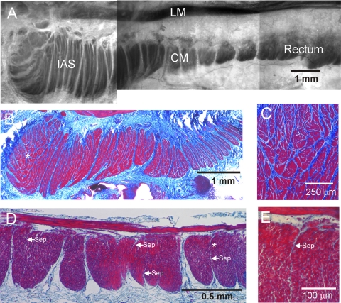
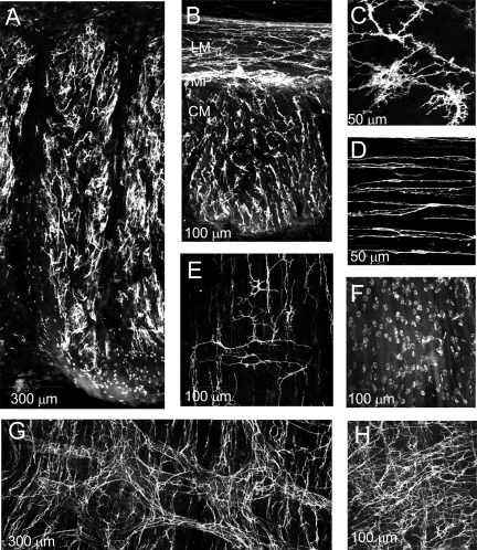
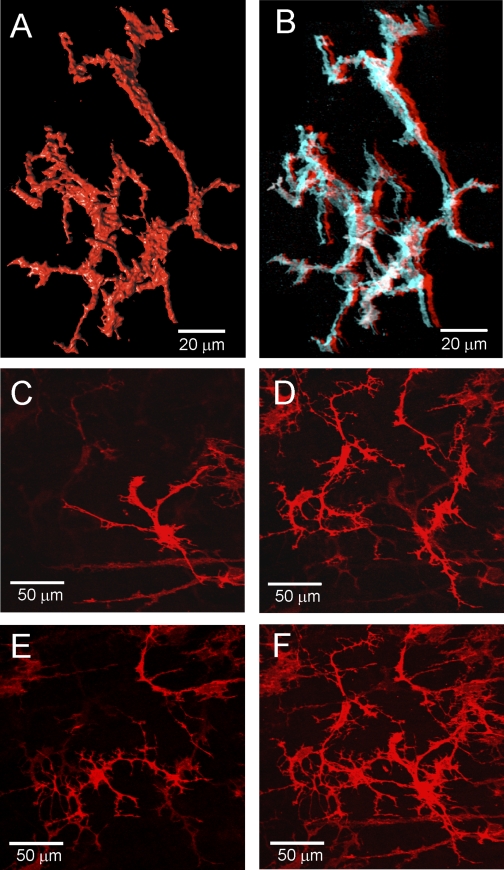
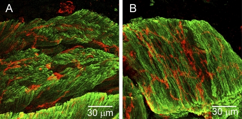
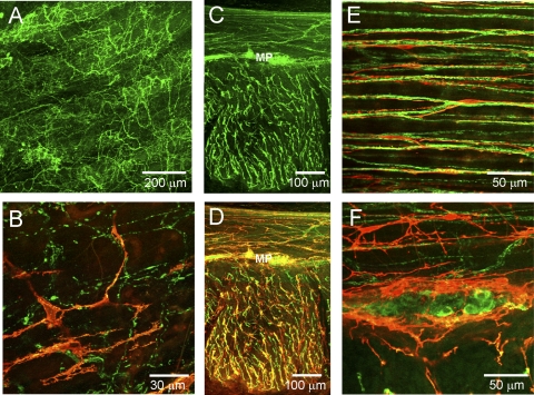
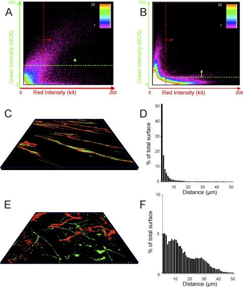
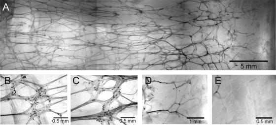
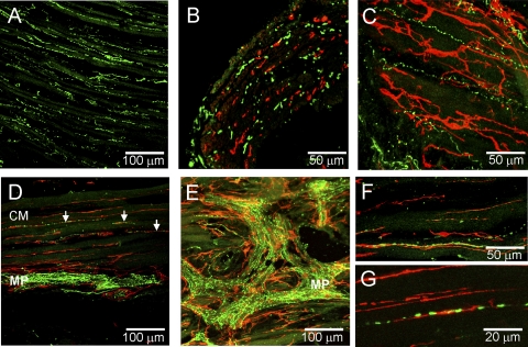
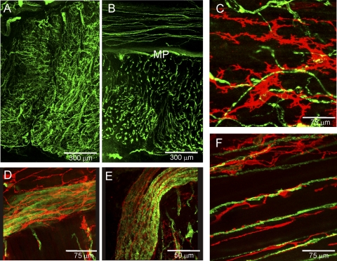
The morphology of interstitial cells of Cajal (ICC) in the circular muscle layer of the cynomolgus monkey internal anal sphincter (IAS) and rectum and their relationship to sympathetic and nitrergic nerves were compared by dual-labeling immunohistochemistry. Contractile studies confirmed that nitrergic nerves participate in neural inhibition in both regions whereas sympathetic nerves serve as excitatory motor nerves only in the IAS. Muscle bundles extended from myenteric to submucosal edge in rectum but in the IAS bundles were further divided into "minibundles" each surrounded by connective tissue. Dual labeling of KIT and smooth muscle myosin revealed KIT-positive stellate-shaped ICC (ICC-IAS) within each minibundle. In the rectum intramuscular ICC (ICC-IM) were spindle shaped whereas stellate-shaped ICC were located at the myenteric surface (ICC-MY). ICC were absent from both the myenteric and submucosal surfaces of the IAS. Nitrergic nerves (identified with anti-neuronal nitric oxide synthase antibodies or NADPH diaphorase activity) and sympathetic nerves (identified with anti-tyrosine hydroxylase antibody) each formed a plexus at the myenteric surface of the rectum but not the IAS. Intramuscular neuronal nitric oxide synthase- and tyrosine hydroxylase-positive fibers were present in both regions but were only closely associated with ICC-IM in rectum. Minimal association was also noted between ICC-IAS and cells expressing the nonspecific neuronal marker PGP9.5. In conclusion, the morphology of rectal ICC-IM and ICC-MY is similar to that described elsewhere in the gastrointestinal tract whereas ICC-IAS are unique. The distribution of stellate-shaped ICC-IAS throughout the musculature and their absence from both the myenteric and submucosal surfaces suggest that ICC-IAS may serve as pacemaker cells in this muscle whereas their limited relationship to nerves suggests that they are not involved in neuromuscular transmission. Additionally, the presence of numerous minibundles, each containing both ICC-IAS and nerves, suggests that this muscle functions as a multiunit type muscle.


Figures

Fig. 1.
Fig. 1.
Morphological features of the monkey rectoanal region. A: composite image of a thick cross section from the rectoanal region; smooth muscle is contrasted with toluidine blue staining. The longitudinal (LM) and circular (CM) muscle layers can be seen separated by connective tissue. Prominent septal structures (white) are also apparent extending from the myenteric to the submucosal edge of the circular muscle separating this layer into bundles. The pinning procedure used for this image has exaggerated the gap between longitudinal and circular muscle layers. A more representative separation can be seen in D. BE: thin (3 μm) cross sections of the internal anal sphincter (IAS) and rectum stained with Masson's trichrome to visualize both smooth muscle (red/purple) and connective tissue (blue). B: low magnification image of the IAS again showing prominent septal structures extending from myenteric (top) to submucosal (bottom) edge. Additional septal structures are apparent further separating the muscle into “minibundles.” C: higher magnification image of the region marked with an asterisk in B showing numerous minibundles (red) separated by connective tissue septa (blue). D: lower magnification image of rectum showing division of the circular muscle layer into compact bundles extending from the myenteric to the submucosal edge and separated by connective tissue septa. A few additional smaller septa can be seen penetrating for variable distances into muscle bundles (arrows labeled “sep”). The region marked with an asterisk in D is shown at higher magnification in E. Cells can be seen between longitudinal and circular muscle layers and these likely include both neurons and interstitial cells of Cajal (ICC) of the myenteric plexus.
Fig. 2.
Fig. 2.
Immunohistochemical labeling of KIT-positive cells in the monkey rectoanal region. KIT-positive ICC (white) are seen distributed throughout the IAS (A) and rectum (B) in these lower magnification (×10) thick cross sections. B: both the longitudinal and circular muscle layers can be seen in this image. Labeling is most dense in the myenteric plexus (MP) region between muscle layers. Higher magnification images of transverse sections show stellate-shaped ICC-IAS in the IAS (C) and spindle-shaped intramuscular ICC in the rectum (D). EH: images showing the distribution of KIT-positive cells at the myenteric and submucosal surfaces of the IAS and rectum in a modified whole mount preparation. Stellate-shaped ICC can be seen at both the submucosal (E, ICC-SM) and myenteric (G and H, ICC-MY) surface of the rectum whereas only small rounded mast cells were observed at the myenteric (F) and submucosal (A) surfaces of the IAS. Optical section thickness: A, 26 μm; B, 17 μm; C, 6 μm; D, 3.75 μm; E, 4 μm; F, 13 μm; G, 18 μm; H, 12 μm. Additional images from modified whole mounts can be seen in Supplemental Figs. S1 and S2.
Fig. 3.
Fig. 3.
Three-dimensional images of ICC in the monkey IAS. A: 3-dimensional (3D) surface reconstruction of a small cluster of ICC-IAS with characteristic branching processes. Image generated from 77 confocal slices (0.15 μm/slice) taken from a cross-sectional preparation (×100). Supplemental Fig. S4 shows rotation of these cells. B: anaglyph of cells shown in A. To view in 3D, red (left eye) and cyan (right eye) glasses are required (see materials and methods). Supplemental Fig. S5 shows anaglyph of a different cluster of cells. CF: lower magnification images (×40) of ICC-IAS within confocal stack (27 slices, 0.25 μm/slice) from a transverse section. CE: 3 different optical sections (each a 1.25-μm stack) selected from top (C), middle (D), and bottom (E) of stack. F: complete confocal stack (13.5 μm thick). A different transverse section taken at lower magnification is shown as an anaglyph in Supplemental Fig. S6 and a movie in Supplemental Video S7.
Fig. 4.
Fig. 4.
Dual labeling of ICC-IAS and smooth muscle cells (SMC) with anti-KIT (red) and anti-SM myosin (green) antibodies. Thick (100 μm) cryostat cross sections of the IAS are shown. SMC were usually oriented vertically (A) but in some cases bundles fell to one side during mounting, allowing a transverse view of SMC and ICC within a 100-μm section of muscle (B). Optical section thickness: A, 3.5 μm; B, 5.75 μm.
Fig. 5.
Fig. 5.
Dual labeling of ICC and nitrergic nerves with anti-KIT (red) and anti-neuronal nitric oxide synthase (nNOS; green) antibodies in the IAS (A and B) and rectum (CF). A: distribution of nNOS-positive cells through the thickness of the IAS in a thick transverse section. B: dual labeling of a thick transverse section of IAS showing marked differences in the overall distribution of nNOS and KIT-positive cells. C and D: 2 images of the same thick cross section of rectal muscle. C: distribution of nNOS positive cells. D: dual image of nNOS and KIT-positive cells at low power (×20) reveals similar distribution of NOS fibers and ICC (yellow). E: high-magnification image of transverse section of circular muscle showing close association of KIT and nNOS-positive cells. F: high-magnification image of a myenteric ganglia containing nNOS-positive cell bodies surrounded by KIT-positive cells. Optical section thickness: A, 18 μm; B, 2 μm; C, 23 μm; D, 23 μm; E, 3.75 μm; F, 11 μm.
Fig. 6.
Fig. 6.
Colocalization and apposition of NOS-labeled and KIT-labeled structures in the IAS and rectum. A and B: plots showing the number of pixels (see spectrum scale bar) averaged throughout the stack at different intensities of red (x-axis) and green (y-axis) in rectum (A) and IAS (B). The quadrant above the green line and to the right of the red line indicates pixels considered to contain both KIT (red) and NOS (green) signals (i.e., sites of colocalization). Greater colocalization of signals is seen in rectum than in IAS. C and E: 3D surface reconstructions (see Ref. 30) of KIT and NOS labeling in IAS (C) and rectal (E) images. D and F: the distance between surface reconstructions was used to obtain the values plotted in histograms D (IAS) and F (rectum) that show the percent of total KIT and NOS surface triangles (y-axis) with minimum distances ranging from 1 to 50 μm (x-axis) in IAS (D) and rectum (F).
Fig. 7.
Fig. 7.
Distribution of NADPH diaphorase-positive enteric nerves along the myenteric surface of a modified whole mount preparation. A: composite image of the overall distribution of cells from the anal verge (AV, right edge) to the proximal rectum (left edge). BD: higher magnification images of NADPH diaphorase positive cells located ∼3 cm (B), 2 cm (C), and 0.7 cm (D) from the AV. E: at 0.4 cm labeling is virtually absent.
Fig. 8.
Fig. 8.
Dual labeling of ICC and sympathetic nerves with anti-KIT (red) and anti-tyrosine hydroxylase (TH, green) antibodies in the IAS (AC) and rectum (DG). A: distribution of TH-positive cells across the thickness of the IAS in a transverse cryosection. Dual labeling of ICC and TH-positive cells in cross section (B) and transverse sections (C) of the IAS. Note general lack of association between KIT-positive cells and TH-positive cells (B and C). D: dual labeling of rectal transverse section reveals a dense plexus of TH-positive cells within the myenteric plexus (MP) of the CM layer and with a few varicose fibers within the CM (arrows). ICC can be seen surrounding TH-positive cells in the plexus region (MP) and closely associated with TH-positive cells within the musculature. E: transverse section through a myenteric ganglia (MP) showing TH-positive cells along with underlying KIT-positive cells. F and G: higher magnification images of ICC-IM and TH-positive fibers showing intimate association. Optical section thickness: A, 14 μm; B, 6 μm; C, 11 μm; D, 6.5 μm; E, 8 μm; F, 8 μm; G, 4.5 μm.
Fig. 9.
Fig. 9.
Dual labeling of ICC and nerves with anti-KIT (red) and anti-PGP9.5 (green) antibodies in the IAS and rectum. Overall distribution of PGP9.5-positive cells in the IAS (A) and rectum (B). CF: dual-labeling images from IAS and rectum. Very limited overlap of KIT and PGP9.5-positive cells is seen in the IAS (C). D and E: nerve trunks identified in the rectal myenteric region with PGP9.5 can be seen surrounded (D) and intertwined (E) with KIT-positive cells. Additional anaglyph images of ICC in nerve trunks are provided as supplemental data (see Supplemental Fig. S3). F: KIT-positive cells are closely aligned with PGP9.5-positive cells in the rectum. Optical section thickness: A, 32 μm; B, 25 μm; C, 2.5 μm; D, 5 μm; E, 7.5 μm; F, 9 μm.

Similar articles

Cited by

References

    1. Baumgarten HG, Holstein AF, Stelzner F. [Differences in the innervation of the large intestine and the internal sphincter of the anus in mammals and humans]. Verh Anat Ges 66: 43–47, 1971 - PubMed
    1. Bharucha AE. Pelvic floor: anatomy and function. Neurogastroenterol Motil 18: 507–519, 2006 - PubMed
    1. Brading AF, Ramalingam T. Mechanisms controlling normal defecation and the potential effects of spinal cord injury. Prog Brain Res 152: 345–358, 2006 - PubMed
    1. Bridgewater M, MacNeil HF, Brading AF. Regulation of tone in pig urethral smooth muscle. J Urol 150: 223–228, 1993 - PubMed
    1. Burns AJ, Herbert TM, Ward SM, Sanders KM. Interstitial cells of Cajal in the guinea-pig gastrointestinal tract as revealed by c-Kit immunohistochemistry. Cell Tissue Res 290: 11–20, 1997 - PubMed

Publication types

Substances

LinkOut - more resources