. 2010 Mar;137(6):963-74.
doi: 10.1242/dev.044719. Epub 2010 Feb 11.

The HMX/NKX homeodomain protein MLS-2 specifies the identity of the AWC sensory neuron type via regulation of the ceh-36 Otx gene in C. elegans

Affiliations

The HMX/NKX homeodomain protein MLS-2 specifies the identity of the AWC sensory neuron type via regulation of the ceh-36 Otx gene in C. elegans

Kyuhyung Kim et al. Development. 2010 Mar.

Abstract

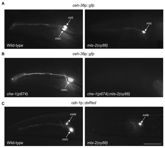
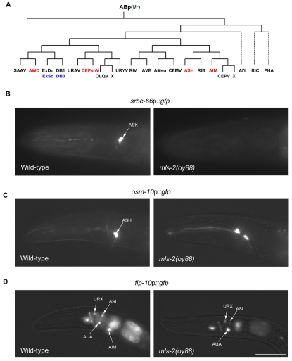
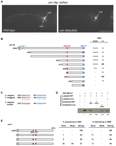
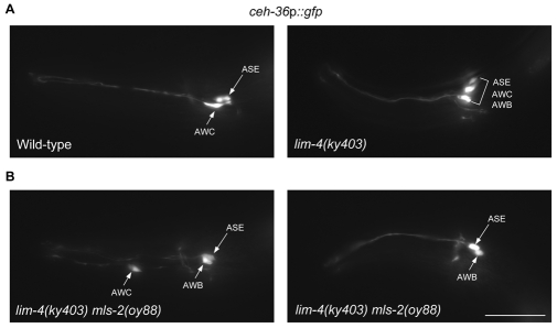
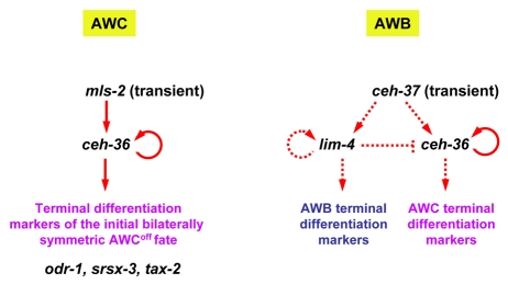
The differentiated features of postmitotic neurons are dictated by the expression of specific transcription factors. The mechanisms by which the precise spatiotemporal expression patterns of these factors are regulated are poorly understood. In C. elegans, the ceh-36 Otx homeobox gene is expressed in the AWC sensory neurons throughout postembryonic development, and regulates terminal differentiation of this neuronal subtype. Here, we show that the HMX/NKX homeodomain protein MLS-2 regulates ceh-36 expression specifically in the AWC neurons. Consequently, the AWC neurons fail to express neuron type-specific characteristics in mls-2 mutants. mls-2 is expressed transiently in postmitotic AWC neurons, and directly initiates ceh-36 expression. CEH-36 subsequently interacts with a distinct site in its cis-regulatory sequences to maintain its own expression, and also directly regulates the expression of AWC-specific terminal differentiation genes. We also show that MLS-2 acts in additional neuron types to regulate their development and differentiation. Our analysis describes a transcription factor cascade that defines the unique postmitotic characteristics of a sensory neuron subtype, and provides insights into the spatiotemporal regulatory mechanisms that generate functional diversity in the sensory nervous system.


Figures

Fig. 1.
Fig. 1.
The C. elegans oy88 mutation affects gene expression in the AWC neurons. (A,B) Expression of a ceh-36p::gfp fusion gene is abolished in the AWC neurons in oy88 mutants in a wild-type (A) or che-1(p674) mutant (B) background. Mutations in the che-1 zinc-finger transcription factor abolish ceh-36p::gfp expression in the ASE neurons (Chang et al., 2003). (C) Expression of an odr-1p::dsRed transgene is affected in the AWC neurons in oy88 mutants. Anterior is to left. Scale bar: 10 μm.
Fig. 2.
Fig. 2.
oy88 is an allele of the mls-2 Hmx/Nkx homeobox transcription factor gene. (A) Genomic structure of C. elegans mls-2 showing the molecular nature of the lesions in mls-2 alleles examined in this work. Exons are indicated by boxes, introns by lines. (B) Alignment of the homeodomains of the indicated proteins, including C. elegans MLS-2. Residues that are identical in at least two proteins are in red. The residue mutated in oy88 is indicated. The Q50 residue in the third helix that is crucial for conferring DNA-binding specificity is underlined. The C-terminal HMX motif that is shared among HMX, but not NKX, proteins is boxed.
Fig. 3.
Fig. 3.
mls-2 is expressed transiently in, and acts in, the AWC neurons to regulate gene expression. (A) Expression of odr-1p::dsRed (left) and a gfp-tagged mls-2 gene driven under its own regulatory sequences (middle) in wild-type L1 larvae. Merged image is on the right. The AWC neurons were identified by their position and odr-1p::dsRed expression. (B) Correlation of the presence of mls-2 rescuing sequences with wild-type ceh-36p::gfp expression in che-1(p674); mls-2(oy88) mutants carrying stably integrated copies of the ceh-36p::gfp fusion gene. mls-2 rescuing sequences in the C39E6 cosmid were present on unstable extrachromosomal arrays that also contain odr-1p::dsRed sequences. The presence of mls-2 sequences in the AWC neurons was monitored by odr-1p::dsRed expression. AWCL, left; AWLR, right. (C) Correlation of the presence of mls-2 rescuing sequences with wild-type AWC neuronal morphology in che-1(p674); mls-2(oy88) mutants carrying stably integrated copies of the ceh-36p::gfp fusion gene. The presence of mls-2 sequences in the AWC neurons was monitored as described in B. (D) An AWC neuron in che-1(p674); mls-2(oy88) animals containing wild-type mls-2 sequences as monitored by the expression of odr-1p::dsRed (left) exhibits a truncated dendrite (arrowhead). The truncated dendrite is also visualized via expression of ceh-36p::gfp (right). Scale bars: 5 μm in A: 10 μm in D.
Fig. 4.
Fig. 4.
MLS-2 regulates the differentiation of multiple ABp(l/r)paa-derived cell types. (A) Lineage diagram of ABp(l/r)-derived cells (not to temporal scale) (see Sulston et al., 1983). Left/right (l/r) asymmetric cells derived from ABpr are shown in blue. Cells affected in mls-2 mutants are in red. Dotted lines indicate lineages for which not all cell divisions are shown. (B) Expression of the ASK marker srbc-66p::gfp (Kim et al., 2009) is abolished in mls-2(oy88) mutants. (C) The ASH sensory neuronal marker osm-10p::gfp (Hart et al., 1999) is ectopically expressed in mls-2(oy88) mutants. (D) Expression of flp-10p::gfp (Kim and Li, 2004) is abolished in the AIM interneurons in mls-2(oy88) animals. Scale bar: 10 μm.
Fig. 5.
Fig. 5.
MLS-2 and CEH-36 interact directly with ceh-36 upstream regulatory sequences to initiate and maintain ceh-36 expression, respectively. (A) CEH-36 autoregulates to maintain its own expression in the AWC neurons. Forty-seven percent of ceh-36(ky640) animals failed to express a ceh-36p::dsRed transgene in the AWC neurons. Expression in the ASE neurons was unaffected. n=66 animals. Scale bar: 10 μm. (B) The extent of ceh-36 upstream regulatory sequences driving gfp expression in the different constructs. The numbering is relative to the initiator ATG. Motifs that interact directly with CEH-36 and MLS-2 are indicated by red and blue boxes, respectively. The CHE-1 binding site is indicated by a green box (Etchberger et al., 2007). Mutated motifs are indicated by an X; the sequences of the mutated motifs are shown in C. The strength of expression is indicated by the number of + symbols (−, no expression detected; ND, not determined). Note that the effects of mutations in the MLS-2 binding motif on transgene-driven gene expression in wild-type AWC neurons can only be observed in the absence of a functional CEH-36 binding site in both L1 larvae and adults. Larval expression driven by sequences containing a functional CEH-36 binding site, but mutated MLS-2 binding site, is likely to be due to the temporal overlap in mls-2 and ceh-36 expression in newly hatched animals. At least 50 animals from two independent transgenic lines carrying each construct were examined. (C) Conservation of the CEH-36 (red) and MLS-2 (blue) binding sites between C. elegans and C. briggsae. Asterisks indicate identical nucleotides. Point mutations in the predicted binding sites analyzed (in B and D) are underlined. (D) Binding of bacterially expressed GST-MLS-2 fusion protein to the predicted MLS-2 binding site upstream of ceh-36 as examined by EMSA. Competition was performed with the indicated excess of unlabeled wild-type or mutant MLS-2 binding sequences shown in C. Numbers below each lane in the gel indicate relative band intensities, with the intensity of the band representing GFP-MLS-2 bound to wild-type sequences set at 1. (E) Mutation of two predicted CEH-36 binding sites (red boxes; mutation indicated by X) in odr-1 upstream regulatory sequences affects odr-1 expression in the AWC neurons. At least 50 animals from two independent transgenic lines carrying each construct were examined.
Fig. 6.
Fig. 6.
mls-2 is not required to drive misexpression of ceh-36 in the AWB neurons in lim-4 mutants. (A) ceh-36p::gfp is misexpressed in the AWB neurons in 100% of lim-4(ky403) mutants. n=50. (B) ceh-36p::gfp expression is retained in the AWB neurons in lim-4(ky403) mls-2(oy88) double mutants. ceh-36p::gfp expression in a mispositioned AWC neuron (left) and in the ASE and AWB neurons (left and right) is shown in lim-4 mls-2 mutants. All lim-4 mls-2 mutants retained ceh-36p::gfp expression in the AWB neurons; n=50. Anterior is to left. Scale bar: 10 μm.
Fig. 7.
Fig. 7.
Models of the developmental regulatory cascades that specify AWC and AWB neuronal identities in C. elegans. In the AWC neurons (left), transient expression of the MLS-2 HMX/NKX transcription factor directly initiates expression of the ceh-36 Otx homeobox gene. CEH-36 maintains expression by autoregulation via direct interaction with its upstream regulatory sequences, and also directly regulates the expression of terminal differentiation genes, including odr-1, srsx-3 and, possibly, tax-2 (Koga and Ohshima, 2004), to specify the initial, bilaterally symmetric AWCOFF fate. In the AWB neurons (right), transient expression of the CEH-37 OTX homeodomain protein initiates expression of the lim-4 LIM-homeobox gene. LIM-4 maintains expression by autoregulation and promotes expression of AWB terminal differentiation genes. CEH-37 may also be able to promote expression of ceh-36 in the AWB neurons, but this expression is normally repressed by LIM-4. In the absence of LIM-4 function, AWB-specific genes are not expressed and the expression of ceh-36 is derepressed, resulting in the expression of AWC fate. Dotted lines indicate regulation that might be direct or indirect.

Similar articles

Cited by

References

    1. Alenina N., Bashammakh S., Bader M. (2006). Specification and differentiation of serotonergic neurons. Stem Cell Rev. 2, 5-10 - PubMed
    1. Alon U. (2007). Network motifs: theory and experimental approaches. Nat. Rev. Genet. 8, 450-461 - PubMed
    1. Altun-Gultekin Z., Andachi Y., Tsalik E. L., Pilgrim D., Kohara Y., Hobert O. (2001). A regulatory cascade of three homeobox genes, ceh-10, ttx-3 and ceh-23 controls cell fate specification of a defined interneuron class in C. elegans. Development 128, 1951-1969 - PubMed
    1. Amendt B. A., Sutherland L. B., Russo A. F. (1999). Transcriptional antagonism between Hmx1 and Nkx2.5 for a shared DNA-binding site. J. Biol. Chem. 274, 11635-11642 - PubMed
    1. Bao Z., Murray J. I., Boyle T., Ooi S. L., Sandel M. J., Waterston R. H. (2006). Automated cell lineage tracing in Caenorhabditis elegans. Proc. Natl. Acad. Sci. USA 103, 2707-2712 - PMC - PubMed

Publication types

MeSH terms