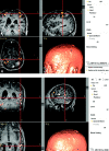
The evolution of clinical functional imaging during the past 2 decades and its current impact on neurosurgical planning
- PMID: 20150316
- PMCID: PMC7964140
- DOI: 10.3174/ajnr.A1845
The evolution of clinical functional imaging during the past 2 decades and its current impact on neurosurgical planning
Abstract
BOLD fMRI has, during the past decade, made a major transition from a purely research imaging technique to a viable clinical technique used primarily for presurgical planning in patients with brain tumors and other resectable brain lesions. This review article briefly examines the history and evolution of clinical functional imaging, with particular emphasis on how the use of BOLD fMRI for neurosurgical planning has changed during the past 2 decades. Even more important, this article describes the many published studies during that same period that have examined the overall clinical impact that BOLD and DTI have made on surgical planning.
Figures


References
-
- Ogawa S, Lee TM, Nayak AS, et al. . Oxygenation-sensitive contrast in magnetic resonance image of rodent brain at high magnetic fields. Magn Reson Med 1990;14:68–78 - PubMed
-
- Ogawa S, Lee TM. Magnetic resonance imaging of blood vessels at high fields: in vivo and in vitro measurements and image simulation. Magn Reson Med 1990;16:9–18 - PubMed
-
- Belliveau JW, Kennedy DN, Jr, McKinstry RC, et al. . Functional mapping of the human visual cortex by magnetic resonance imaging. Science 1991;254:716–19 - PubMed
-
- Chakraborty A, McEvoy AW. Presurgical functional mapping with functional MRI. Curr Opin Neurol 2008;21:446–51 - PubMed
Publication types
MeSH terms
LinkOut - more resources
Full Text Sources
Medical
Miscellaneous
