. 2010 Apr;16(4):720-31.
doi: 10.1261/rna.1963810. Epub 2010 Feb 11.

Role of miR-34c microRNA in the late steps of spermatogenesis

Affiliations

Role of miR-34c microRNA in the late steps of spermatogenesis

Frantz Bouhallier et al. RNA. 2010 Apr.

Abstract

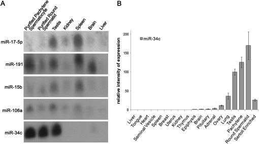
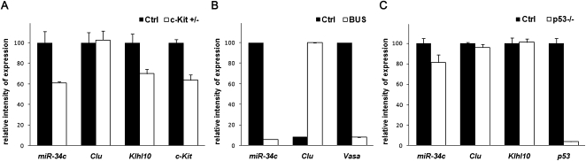
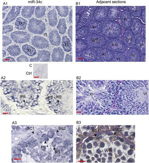
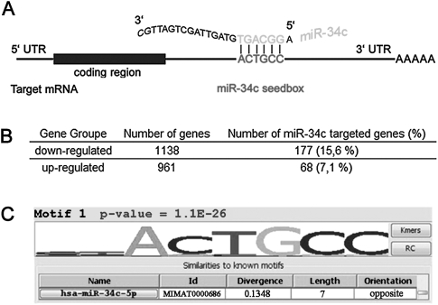
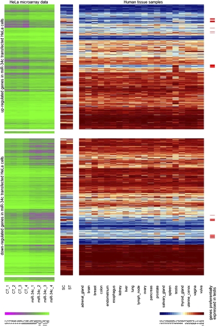
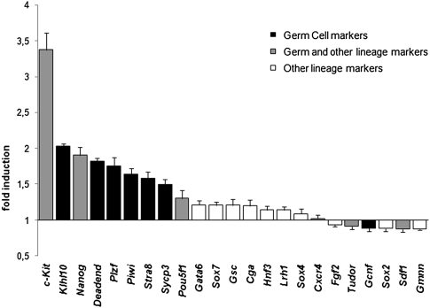
Spermatogenesis is a cyclic process in which diploid spermatogonia differentiate into haploid spermatozoa. This process is highly regulated, notably at the post-transcriptional level. MicroRNAs (miRNAs), single-stranded noncoding RNA molecules of about 20-25 nucleotides, are implicated in the regulation of many important biological pathways such as proliferation, apoptosis, and differentiation. We wondered whether miRNAs could play a role during spermatogenesis. The miRNA expression repertoire was tested in germ cells, and we present data showing that miR-34c was highly expressed only in these cells. Furthermore, our findings indicate that in male gonads, miR-34c expression is largely p53 independent in contrast to previous results showing a direct link in somatic cells between the miR-34 family and this tumor suppressor protein. In order to identify target genes involved in germinal lineage differentiation, we overexpressed miR-34c in HeLa cells, analyzed the transcriptome of these modified cells, and noticed a shift of the expression profile toward the germinal lineage. Recently, it has been shown that exogenous expression of Ddx4/Vasa in embryonic chicken stem cells (cESC) induces cESC reprogramming toward a germ cell fate. When we simultaneously expressed miR-34c in such cells, we could detect an up-regulation of germ cell-specific genes whereas the expression of other lineage specific markers remained unchanged. These data suggest that miR-34c could play a role by enhancing the germinal phenotype of cells already committed to this lineage.


Figures

FIGURE 1.
FIGURE 1.
miR-34c expression profile in different mouse tissues. (A) Northern blot analysis of miR-34c, miR-106a, miR-15b, miR-191, and miR-17-5p expression in different tissues. Thirty micrograms of total RNA were loaded in each well and hybridized with the indicated probes. (B) RT-qPCR analysis of miR-34c expression in various tissues. A value of 100 was given to the miR-34c relative expression in testis. Two different RT reactions were carried out three times and used to calculate the mean and standard error (SEM).
FIGURE 2.
FIGURE 2.
miR-34c is expressed preferentially in germ cells. (A) miR-34c expression in adult mouse c-Kit ± testis. miR-34c, Clusterin (Sertoli marker), Klhl10 (germ cell marker), and c-Kit RNA expressions were tested by RT-qPCR. (B) miR-34c expression in busulfan-treated adult mouse testis. miR-34c, Clusterin (Sertoli marker) and Vasa (germ cell marker) RNA expressions were tested by RT-qPCR. (C) miR-34c expression in p53-deficient mouse testis. miR-34c, Clusterin (Sertoli marker), Klhl10 (germ cell marker), and p53 RNA expressions were tested by RT-qPCR. For each experiment, a value of 100 was given to the relative gene expression in the wild-type mouse testis (Ctrl). Three different RT reactions were carried out three times and used to calculate the mean and standard error (SEM).
FIGURE 3.
FIGURE 3.
Localization of miR-34c by in situ hybridization of adult mouse seminiferous tubule sections. (Section A1) A significant signal was observed in all tubules and was more intense in late stages (IX and XI) of the seminiferous epithelium. No signal was noticed in interstitial tissue (IT). (Section A2) The expression of miR-34c was localized in meiotic cells: young pachytene spermatocytes (yPS), late pachytene spermatocytes (LPS), and in round spermatids (RS). (Section A3) No signal was observed in Sertoli cells (SC). Two Sertoli cells (SC1 and SC2) are shown on the section treated with miR-34c probe and on the control adjacent section. Note that when the nucleus of a SC is seen on a section, only a small part of it is present on the adjacent section. (Section C) Background in the absence of the miR-34c-specific probe. The stages of the seminiferous epithelium cycle (roman numerals) were identified from adjacent sections B1, B2, and B3. Adjacent sections B1 and B2) were stained with PAS and hematoxylin. Adjacent section B3 was incubated with an antivimentin antibody (a somatic cell marker) and stained with PAS and hematoxylin. Magnification: sections A1, B1, and C, X100; sections A2 and B2, ×400; and sections A3 and B3, ×1000. Bars in sections A1, B1, and C represent 50 μm. Bars in sections A2, B2, A3, and B3 represent 10 μm.
FIGURE 4.
FIGURE 4.
Sites complementary to the miR-34c targeted sequence are enriched in the 3′UTRs of down-regulated transcripts. (A) Schematic representation of miR-34c base pairing with its targets. (B) miRGen results: miRGen (http://www.diana.pcbi.upenn.edu/cgi-bin/miRGen/v3/Targets.cgi) is a tool that provides a list of genes potentially recognized by a miRNA. We compared the miR-34c targeted gene list provided by miRGen with our experimental lists (DNA arrays on miR-34c overexpressing cells) of up- and down-regulated genes. The down-regulated gene list harbored more miR-34c regulated genes in comparison with up-regulated genes (15.6% versus 7.1%). (C) Allegro results: overrepresented motifs in the 3′UTRs of down-regulated genes. We submitted the miR-34c down- and up-regulated gene lists to the allegro software (http://acgt.cs.tau.ac.il/allegro/). Allegro is a software tool for discovering motifs statistically overrepresented in gene 3′UTR. The only noticeable motif highlighted by this analysis was a sequence corresponding to the miR-34c seedbox (ACTGCC) in the down-regulated gene list, whereas no particular motif was recognized in the up-regulated gene list.
FIGURE 5.
FIGURE 5.
The expression of the up- and downregulated genes in purified germ cells and human tissue samples. A color-coded diagram (heatmap) of the up- and down-regulated genes is shown. Each column corresponds to a sample. Each line represents an Operon probe and its log2-transformed intensity signal: from pink (low signal) to green (high signal), as indicated in the bottom-left scale bar in the four control HeLa cells (CT_1–CT_4) and in the HeLa cells overexpressing miR-34c (miR-34c_1–miR-34c_4). Log2-transformed intensity signals of the affymetrix probe sets: from blue (low signal) to red (high signal), as indicated in the bottom-right scale bar, corresponding to the same gene were also displayed. These samples, hybridized on the Affymetrix GeneChip Human Genome U133 Plus 2.0 microarray, correspond to purified pachytene spermatocytes (SC) and round spermatids (ST), as well as 20 healthy human tissues including adrenal gland, brain, breast, colon, endometrium, esophagus, kidney, liver, lung, lymph node, ovary, pancreas, salivary gland, spleen, testis, thyroid gland, uterine cervix, vagina, and vulva. Probes showing a preferential expression in testis are shown with a red bar in the last column (right). Operon probes corresponding to genes not representated by affymetrix probe sets had no intensity signal using this technology (second and fourth sets of probes).
FIGURE 6.
FIGURE 6.
TGIF2 and NOTCH2 are direct targets of miR-34c. (A) We validated TGIF2 down-regulation by miR-34c in HeLa transfected cells by RT-qPCR. (B) Tgif2 has a low expression in mouse germ cells but is highly expressed in testis somatic cells, in contrast with miR-34c. Tgif2 expression was tested in bulsulfan-treated mouse testis (BUS), pachytene spermatocytes (PACH), round spermatids (RS), and in control testis (Ctrl). RT-qPCR showed that Tgif2 is highly expressed in bulsulfan-treated mouse testis (depleted in germ cells) when compared with the control untreated testis and to purified germ cells. For each experiment, a value of 100 was given to the control cells (testis). Three different RT reactions were carried out three times and used to calculate the mean and standard error (SEM). (C) Luciferase assays demonstrated that miR-34c directly down-regulates TGIF2 by targeting the TGIF2 3′UTR. Luciferase reporter constructs containing full-length (WT) and mutated miR-34c seedbox (MUT) 3′UTR of TGIF2 were cotransfected with pre-miR-34c miRNA precursor (miR-34c) or pre-miR miRNA precursor RNA negative control (Ctrl) into HeLa cells. Luciferase activity was measured 48 h after transfection. Mutation in the miR-34c binding site (MUT) resulted in a reduction of the inhibition induced by miR-34c. A value of 100 was given to the control cells. Three different transfections were carried out and luciferase activity was measured twice for each reaction. (D,E,F) The same experiments were undertaken with NOTCH2 and showed similar results.
FIGURE 7.
FIGURE 7.
miR-34c overexpression enhanced the germ profile of cells already engaged in the first steps of the germ lineage. Chicken ES cells overexpressing chicken Vasa homolog (Cvh) and cultured under appropriate conditions differentiate toward the first steps of spermatogenesis. We cotransfected miR-34c in these Cvh overexpressing ES chicken cells cultured under the same conditions and analyzed the expression profile of selected differentiation markers. We analyzed the expression level of these markers by RT-qPCR in the miR-34c & Cvh co-transfected cells versus the Cvh control cells. A value of 1 was given to the expression level of these markers in the Cvh ES cells and the resulting up or down-regulation of these markers was noted on this graph.

Similar articles

Cited by

References

    1. Ambros V. The evolution of our thinking about microRNAs. Nat Med. 2008;14:1036–1040. - PubMed
    1. Bannister SC, Tizard ML, Doran TJ, Sinclair AH, Smith CA. Sexually dimorphic microRNA expression during chicken embryonic gonadal development. Biol Reprod. 2009;81:165–176. - PMC - PubMed
    1. Bartel DP. MicroRNAs: Target recognition and regulatory functions. Cell. 2009;136:215–233. - PMC - PubMed
    1. Baskerville S, Bartel DP. Microarray profiling of microRNAs reveals frequent coexpression with neighboring miRNAs and host genes. RNA. 2005;11:241–247. - PMC - PubMed
    1. Bommer GT, Gerin I, Feng Y, Kaczorowski AJ, Kuick R, Love RE, Zhai Y, Giordano TJ, Qin ZS, Moore BB, et al. p53-mediated activation of miRNA34 candidate tumor-suppressor genes. Curr Biol. 2007;17:1298–1307. - PubMed

Publication types