Review
. 2010 Feb;12(2):199-211.
doi: 10.1093/neuonc/nop022. Epub 2009 Dec 14.

The functional role of Notch signaling in human gliomas

Affiliations
Review

The functional role of Notch signaling in human gliomas

Marie-Thérése Stockhausen et al. Neuro Oncol. 2010 Feb.

Abstract

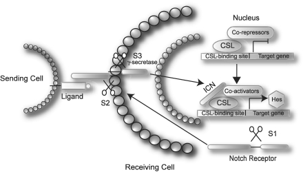
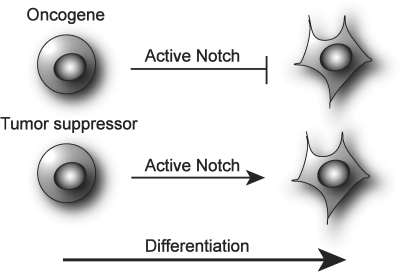
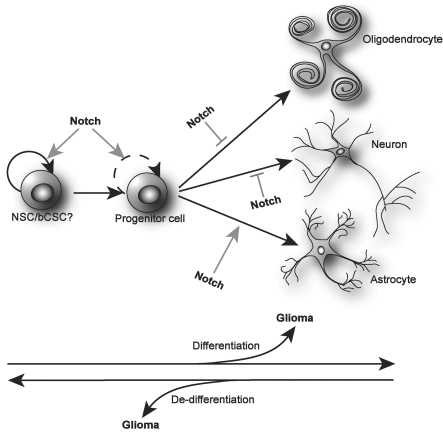
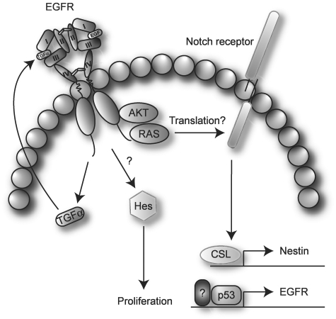
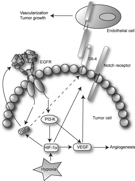
Gliomas are among the most devastating adult tumors for which there is currently no cure. The tumors are derived from brain glial tissue and comprise several diverse tumor forms and grades. Recent reports highlight the importance of cancer-initiating cells in the malignancy of gliomas. These cells have been referred to as brain cancer stem cells (bCSC), as they share similarities to normal neural stem cells in the brain. The Notch signaling pathway is involved in cell fate decisions throughout normal development and in stem cell proliferation and maintenance. The role of Notch in cancer is now firmly established, and recent data implicate a role for Notch signaling also in gliomas and bCSC. In this review, we explore the role of the Notch signaling pathway in gliomas with emphasis on its role in normal brain development and its interplay with pathways and processes that are characteristic of malignant gliomas.


Figures

Fig. 1.
Fig. 1.
Schematic overview of the canonical Notch signaling pathway. The Notch receptors are produced as large proteins that are cleaved (S1) and inserted in the membrane as heterodimers. Upon ligand binding, two consecutive cleavages occur (S2 and S3), which release the ICN. In the nucleus, ICN forms a multimeric protein complex together with CSL and co-activators, and initiates transcription of target genes. See text for further details.
Fig. 2.
Fig. 2.
Notch as an oncogene or tumor suppressor. It is hypothesized that Notch functions as an oncogene in cells where Notch is activated to maintain an undifferentiated phenotype and has to be down-regulated in order for differentiation to occur. In contrast, in cells where Notch activity is required for differentiation to proceed, it is thought to function as a tumor suppressor as its absence maintains cells in an undifferentiated state.
Fig. 3.
Fig. 3.
A simplified view of Notch in NSC and development of gliomas. Notch signaling is important for the self-renewal of NSC, and possibly bCSC. From these stem cells, progenitor cells arise that again will form more and more lineage restricted progenitors. In some of these developmental steps, Notch is involved in the self-renewing process of the progenitor cells. Finally, an active Notch cascade is involved in the terminal differentiation of astrocytes and, conversely, inhibits the final maturation of neurons and oligodendrocytes. The cell of origin for gliomas is currently not known. However, it is possible that gliomas might arise from transforming events in every developmental step from the NSC to mature CNS cells. It is also possible that de-differentiation of more mature cells may lead to gliomagenesis. As such the bCSC might be either the initiator of glioma growth or a result of glioma progression.
Fig. 4.
Fig. 4.
Schematic illustration of Notch and EGFR cross-talk in glioma. Activation of AKT and/or RAS induces expression of Notch, possibly by recruiting existing Notch mRNAs to polysomes and increasing translation. Notch drives the expression of Nestin and EGFR, in part through activation of p53. Signaling through EGFR up-regulates TGFα, which induces expression of Hes and leads to proliferation. The mechanism behind up-regulation of Hes by TGFα is currently not elucidated but has been suggested to occur without Notch activation. See text for further details.
Fig. 5.
Fig. 5.
Potential Notch and EGFR interaction in glioma angiogenesis. Hypoxia and EGFR both induce expression of TGFα, creating an autocrine loop for EGFR activation. Upon EGFR signaling and in response to hypoxia, HIF-1α and VEGF that are involved in angiogenesis are up-regulated. Furthermore, Notch receptors and ligands are induced by HIF-1α and VEGF. Dll-4 expression in glioma tumor cells activates Notch signaling in nearby endothelial cells leading to vascularization and tumor growth. See text for further details.

Comment in

Similar articles

Cited by

References

    1. Stupp R, Mason WP, van den Bent MJ, et al. Radiotherapy plus concomitant and adjuvant temozolomide for glioblastoma. N Engl J Med. 2005;10:987–996. - PubMed
    1. Galli R, Binda E, Orfanelli U, et al. Isolation and characterization of tumorigenic, stem-like neural precursors from human glioblastoma. Cancer Res. 2004;19:7011–7021. - PubMed
    1. Singh SK, Clarke ID, Terasaki M, et al. Identification of a cancer stem cell in human brain tumors. Cancer Res. 2003;18:5821–5828. - PubMed
    1. Bao S, Wu Q, McLendon RE, et al. Glioma stem cells promote radioresistance by preferential activation of the DNA damage response. Nature. 2006;444:756–760. - PubMed
    1. Artavanis-Tsakonas S, Rand MD, Lake RJ. Notch signaling: cell fate control and signal integration in development. Science. 1999;5415:770–776. - PubMed

Publication types

MeSH terms

Substances