. 2010 Apr 9;285(15):11418-26.
doi: 10.1074/jbc.M109.090746. Epub 2010 Feb 11.

Regulation of adhesion dynamics by calpain-mediated proteolysis of focal adhesion kinase (FAK)

Affiliations

Regulation of adhesion dynamics by calpain-mediated proteolysis of focal adhesion kinase (FAK)

Keefe T Chan et al. J Biol Chem. .

Abstract

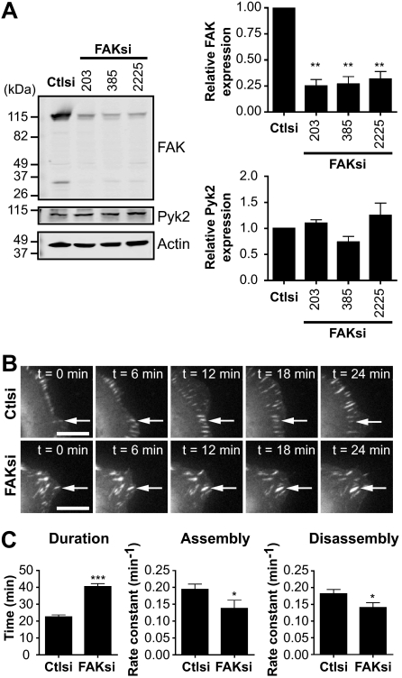
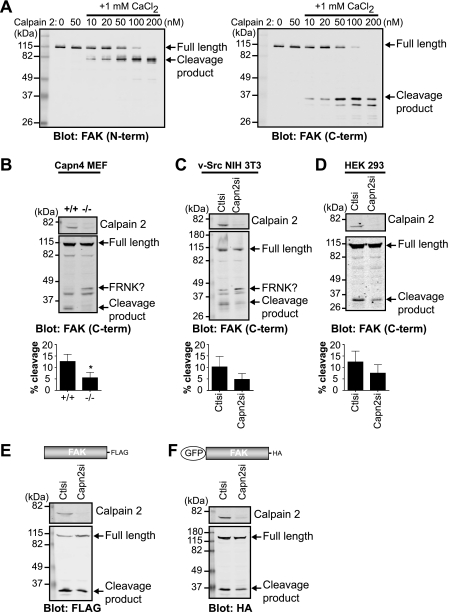
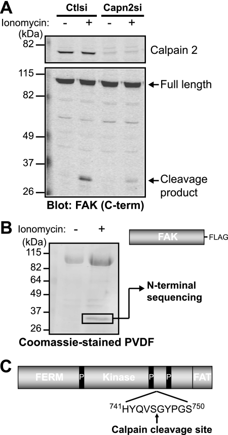
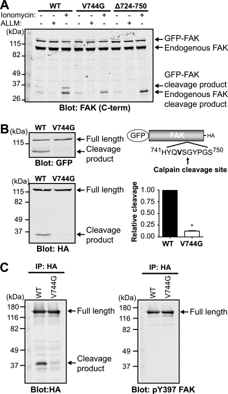
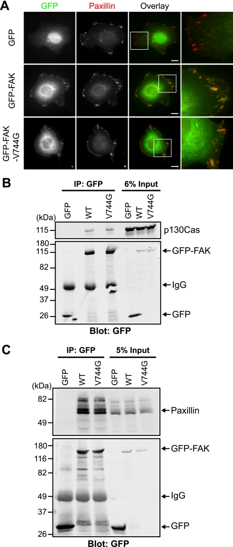
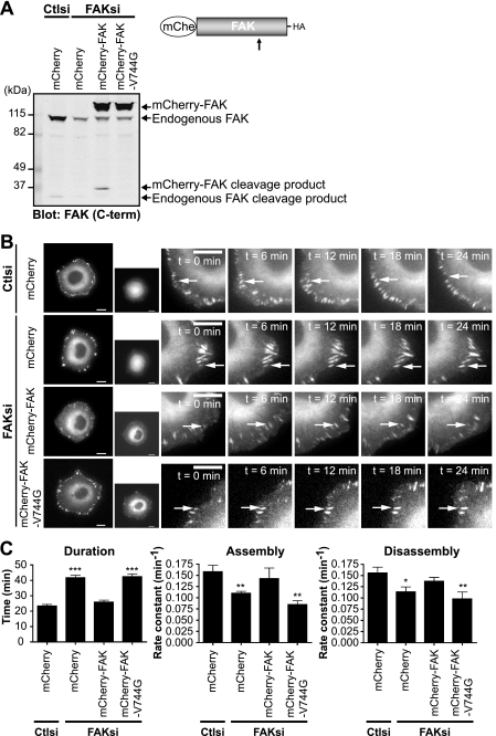
The coordinated and dynamic regulation of adhesions is required for cell migration. We demonstrated previously that limited proteolysis of talin1 by the calcium-dependent protease calpain 2 plays a critical role in adhesion disassembly in fibroblasts (Franco, S. J., Rodgers, M. A., Perrin, B. J., Han, J., Bennin, D. A., Critchley, D. R., and Huttenlocher, A. (2004) Nat. Cell Biol. 6, 977-983). However, little is known about the contribution of other calpain substrates to the regulation of adhesion dynamics. We now provide evidence that calpain 2-mediated proteolysis of focal adhesion kinase (FAK) regulates adhesion dynamics in motile cells. We mapped the preferred calpain cleavage site between the two C-terminal proline-rich regions after Ser-745, resulting in a C-terminal fragment similar in size to the FAK-related non-kinase (FRNK). We generated mutant FAK with a point mutation (V744G) that renders FAK resistant to calpain proteolysis but retains other biochemical properties of FAK. Using time-lapse microscopy, we show that the dynamics of green fluorescent protein-talin1 are impaired in FAK-deficient cells. Expression of wild-type but not calpain-resistant FAK rescues talin dynamics in FAK-deficient cells. Taken together, our findings suggest a novel role for calpain proteolysis of FAK in regulating adhesion dynamics in motile cells.


Figures

FIGURE 1.
FIGURE 1.
FAK regulates adhesion turnover in HEK 293 cells. A, cell lysates from HEK 293 cells transiently transfected with control siRNA (Ctlsi) or FAK siRNA (FAKsi; FAKsi-203, FAKsi-385, FAKsi-2225) were analyzed by immunoblotting and probed for FAK and Pyk2. Actin was probed as a loading control. Immunoblots shown are from one of four separate experiments. Quantification of relative FAK or Pyk2 expression is defined as the ratio of FAK or Pyk2 to the expression in control siRNA. Relative FAK or Pyk2 expression is shown as the mean ± S.E. **, p < 0.01 (by one-way analysis of variance) compared with control siRNA. B, GFP-talin1 was transiently cotransfected with control or FAK (FAKsi-203) siRNA into HEK 293 cells. Cells were plated on fibronectin-coated glass-bottom dishes and analyzed by time-lapse fluorescence microscopy. Time-lapse montages demonstrate representative images of the dynamics of GFP-talin1 over a period of 24 min. Scale bars = 10 μm. Arrows indicate a representative adhesion. Representative movies are shown in supplemental Movies 1 and 2. C, duration was measured as the time elapsed between the appearance and dissolution of an observed adhesion. Rate constants for net adhesion assembly and disassembly were calculated from plots of fluorescence intensities of GFP-talin1 as described under “Experimental Procedures.” Data for each condition are the mean ± S.E. of a total of six cells over three independent experiments. *, p < 0.05; ***, p < 0.001 (by t test) compared with control siRNA.
FIGURE 2.
FIGURE 2.
FAK is cleaved by calpain 2 in vitro and in vivo. A, in vitro calpain cleavage assay of FAK was performed by incubating FAK in cleavage buffer alone or with calpain 2 or with 1 mm CaCl2 and increasing concentrations of calpain 2. Cleavage reactions were analyzed by immunoblotting and probed with anti-FAK antibodies specific to the N-terminal (N-term) and C-terminal (C-term) regions of FAK. Immunoblots shown are from one of two independent experiments. B–D, cell lysates from wild-type (+/+) and calpain 4 knock-out (−/−) mouse embryonic fibroblasts (Capn4 MEF) (B); v-Src-transformed NIH 3T3 control (Ctlsi) and calpain 2 (Capn2si) siRNA fibroblasts (C); and HEK 293 parental, control, and calpain 2 siRNA cells (D) were analyzed by immunoblotting and probed for calpain 2 and FAK. Immunoblots shown are from one of three independent experiments. Quantification of percent cleavage is defined as the ratio of cleaved FAK to total FAK (full-length + cleaved). Percent cleavage is shown as the mean ± S.E. *, p < 0.05 (by t test) compared with the control. E and F, cell lysates from HEK 293 control and calpain 2 siRNA cells transiently transfected with FAK-FLAG (E) or GFP-FAK (F) were analyzed by immunoblotting and probed with anti-calpain 2 and anti-FLAG (E) or anti-HA (F) antibodies. Immunoblots shown are from one of three independent experiments.
FIGURE 3.
FIGURE 3.
Calpain cleaves FAK between the two C-terminal proline-rich regions. A, HEK 293 control (Ctlsi) or calpain 2 (Capn2si) siRNA cells were treated with vehicle (−) or 1 μm ionomycin + 1 mm CaCl2 (+). Cell lysates were analyzed by immunoblotting and probed for calpain 2 and FAK. Immunoblots shown are from one of two independent experiments. C-term, C-terminal. B, FAK-FLAG was transiently transfected into HEK 293 cells, which were treated with vehicle (−) or 1 μm ionomycin + 1 mm CaCl2 (+). Cell lysates were incubated with anti-FLAG beads, and protein complexes were transferred to polyvinylidene difluoride membrane. The 35-kDa band was analyzed by N-terminal sequencing. The Coomassie Blue-stained polyvinylidene difluoride (PVDF) membrane shown is from one of three independent experiments. C, a schematic of FAK domain organization (4.1, ezrin, radixin, moesin (FERM); kinase; focal adhesion targeting (FAT)) denotes the site of calpain cleavage after Ser-745, between the two C-terminal proline-rich (P) regions.
FIGURE 4.
FIGURE 4.
Point mutant of FAK is resistant to calpain proteolysis. A, HEK 293 cells were transfected with wild-type GFP-FAK (WT), GFP-FAK-V744G, or GFP-FAKΔ724–750. Cells were treated with vehicle (−), 50 μg/ml calpain inhibitor N-acetyl-Leu-Leu-Met (ALLM), or 1 μm ionomycin and 10 mm CaCl2. Cell lysates were analyzed by immunoblotting and probed for FAK. The immunoblot shown is from one of three independent experiments. C-term, C-terminal. B, cell lysates from HEK 293 cells transiently transfected with GFP-FAK or GFP-FAK-V744G were analyzed by immunoblotting and probed with anti-GFP and anti-HA antibodies. Immunoblots shown are from one of five independent experiments. Quantification of relative cleavage is defined as the ratio of cleaved GFP-FAK to total GFP-FAK (full-length + cleaved) relative to GFP-FAK. Relative cleavage is shown as the mean ± S.E. *, p < 0.05 (by t test) compared with wild-type GFP-FAK. C, HEK 293 cells were transiently transfected with GFP-FAK or GFP-FAK-V744G. Proteins were immunoprecipitated (IP) with anti-HA antibody, followed by immunoblotting with anti-HA and anti-phospho-Tyr-397 (pY397) FAK antibodies. Immunoblots shown are from one of three independent experiments.
FIGURE 5.
FIGURE 5.
Calpain-resistant FAK localizes to adhesions and retains biochemical properties. A, HEK 293 cells transiently transfected with GFP, GFP-FAK, or GFP-FAK-V744G were cultured on fibronectin-coated coverslips and stained with anti-paxillin antibody. Images shown are representative of three independent experiments. Scale bars = 10 μm. Regions outlined by boxes correspond to magnified images of adhesions. B and C, HEK 293 cells were transiently transfected with GFP, GFP-FAK, or GFP-FAK-V744G. Proteins were immunoprecipitated (IP) with anti-GFP antibody. Cell lysates and co-immunoprecipitations were analyzed by immunoblotting and probed with anti-GFP antibody and anti-p130Cas (B) or anti-paxillin (C) antibody. Immunoblots shown are from one of at least three independent experiments. WT, wild-type GFP-FAK.
FIGURE 6.
FIGURE 6.
Expression of wild-type but not calpain-resistant FAK rescues impaired adhesion turnover in FAK-deficient cells. A, HEK 293 cells were transiently cotransfected with control (Ctlsi) or FAK (FAKsi) siRNA and mCherry (mChe), mCherry-FAK, or mCherry-FAK-V744G. Cell lysates were analyzed by immunoblotting and probed for FAK. The immunoblot shown is from one of three independent experiments. C-term, C-terminal. B, HEK 293 cells stably expressing mCherry, mCherry-FAK, or mCherry-FAK-V744G were transiently cotransfected with control or FAK siRNA and GFP-talin1. Cells were plated on fibronectin-coated glass-bottom dishes and analyzed by time-lapse fluorescence microscopy. Time-lapse montages demonstrate representative images of the dynamics of GFP-talin1 over a period of 24 min. Scale bars = 10 μm. Arrows indicate a representative adhesion. Representative movies are shown in supplemental Movies 3–6. C, duration was measured as the time elapsed between the appearance and dissolution of an observed adhesion. Rate constants for net adhesion assembly and disassembly were calculated from plots of fluorescence intensities of GFP-talin1 as described under “Experimental Procedures.” Data for each condition are the mean ± S.E. from a total of six cells over three independent experiments. *, p < 0.05; **, p < 0.01; ***, p < 0.001 (by t test) compared with control siRNA.

Similar articles

Cited by

References

    1. Ridley A. J., Schwartz M. A., Burridge K., Firtel R. A., Ginsberg M. H., Borisy G., Parsons J. T., Horwitz A. R. (2003) Science 302, 1704–1709 - PubMed
    1. Sieg D. J., Hauck C. R., Schlaepfer D. D. (1999) J. Cell Sci. 112, 2677–2691 - PubMed
    1. Schaller M. D., Hildebrand J. D., Shannon J. D., Fox J. W., Vines R. R., Parsons J. T. (1994) Mol. Cell. Biol. 14, 1680–1688 - PMC - PubMed
    1. Webb D. J., Donais K., Whitmore L. A., Thomas S. M., Turner C. E., Parsons J. T., Horwitz A. F. (2004) Nat. Cell Biol. 6, 154–161 - PubMed
    1. Schober M., Raghavan S., Nikolova M., Polak L., Pasolli H. A., Beggs H. E., Reichardt L. F., Fuchs E. (2007) J. Cell Biol. 176, 667–680 - PMC - PubMed

Publication types