

Multiplexed assessment of the Southwest Oncology Group-directed Intergroup Breast Cancer Trial S9313 by AQUA shows that both high and low levels of HER2 are associated with poor outcome
- PMID: 20150438
- PMCID: PMC2843456
- DOI: 10.2353/ajpath.2010.090711
Multiplexed assessment of the Southwest Oncology Group-directed Intergroup Breast Cancer Trial S9313 by AQUA shows that both high and low levels of HER2 are associated with poor outcome
Abstract
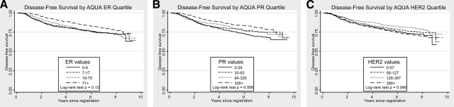
Assessment of key breast cancer tissue biomarkers is often done using nonquantitative methods. We hypothesized that use of continuous analysis of expression with the AQUA method of automated quantitative analysis will provide prognostic information beyond that attainable with conventional methods. A tissue microarray was made from 2123 of 3122 patients accrued to SWOG 9313, in which sequential doxorubicin (A) and cyclophosphamide (C) was compared with combination AC and in which all patients except premenopausal estrogen receptor (ER)-negative patients received tamoxifen. Multiplexed assays of 1) HER2 and estrogen receptor and 2) progesterone receptor (PgR) and p53 were performed on the two slides using the immunofluorescence-based AQUA method of automated quantitative analysis. Both ER and PgR showed unimodal distributions and significantly predicted disease-free survival when tested as continuous variables and adjusted for node status, tumor size, treatment, and menopausal status (P = 0.005 and P < 0.001, respectively). HER2, measured as a continuous variable, showed a biphasic effect on disease-free survival. Both high and low expressers of HER2 have worse outcomes (when low levels are equivalent to that seen in normal breast ducts). In patients who were uniformly treated with AC chemotherapy and tamoxifen (when indicated), both ER and PgR, assessed as continuous variables, were highly prognostic, whereas p53 expression was not. This assay method may provide a new companion diagnostic approach for targeted therapies.
Figures


Similar articles
-
Prognostic significance of progesterone receptor levels in estrogen receptor-positive patients with metastatic breast cancer treated with tamoxifen: results of a prospective Southwest Oncology Group study.J Clin Oncol. 1992 Aug;10(8):1284-91. doi: 10.1200/JCO.1992.10.8.1284. J Clin Oncol. 1992. PMID: 1634918
-
High Ki-67 Expression and Low Progesterone Receptor Expression Could Independently Lead to a Worse Prognosis for Postmenopausal Patients With Estrogen Receptor-Positive and HER2-Negative Breast Cancer.Clin Breast Cancer. 2015 Jun;15(3):204-11. doi: 10.1016/j.clbc.2014.12.007. Epub 2014 Dec 24. Clin Breast Cancer. 2015. PMID: 25600243
-
Clinicopathological factors predicting early and late distant recurrence in estrogen receptor-positive, HER2-negative breast cancer.Breast Cancer. 2016 Nov;23(6):830-843. doi: 10.1007/s12282-015-0649-0. Epub 2015 Oct 14. Breast Cancer. 2016. PMID: 26467036
-
Optimal adjuvant endocrine treatment of ER+/HER2+ breast cancer patients by age at diagnosis: A population-based cohort study.Eur J Cancer. 2018 Feb;90:92-101. doi: 10.1016/j.ejca.2017.11.010. Epub 2017 Dec 21. Eur J Cancer. 2018. PMID: 29274928
-
HER-2/neu and p53 expression versus tamoxifen resistance in estrogen receptor-positive, node-positive breast cancer.J Clin Oncol. 2000 Oct 15;18(20):3471-9. doi: 10.1200/JCO.2000.18.20.3471. J Clin Oncol. 2000. PMID: 11032587
Cited by
-
Breast Cancer Staging: Is TNM Ready to Evolve?J Glob Oncol. 2018 Sep;4:1-3. doi: 10.1200/JGO.17.00004. Epub 2017 Aug 28. J Glob Oncol. 2018. PMID: 30241210 Free PMC article. No abstract available.
-
Spatially multiplexed RNA in situ hybridization to reveal tumor heterogeneity.Nucleic Acids Res. 2020 Feb 20;48(3):e17. doi: 10.1093/nar/gkz1151. Nucleic Acids Res. 2020. PMID: 31853536 Free PMC article.
-
Internodal HER2 heterogeneity of axillary lymph node metastases in breast cancer patients.Bosn J Basic Med Sci. 2019 Aug 20;19(3):242-248. doi: 10.17305/bjbms.2019.3970. Bosn J Basic Med Sci. 2019. PMID: 30957723 Free PMC article.
-
Image analysis of immunohistochemistry is superior to visual scoring as shown for patient outcome of esophageal adenocarcinoma.Histochem Cell Biol. 2015 Jan;143(1):1-9. doi: 10.1007/s00418-014-1258-2. Epub 2014 Aug 26. Histochem Cell Biol. 2015. PMID: 25156293
-
Tumor-associated macrophage, angiogenesis and lymphangiogenesis markers predict prognosis of non-small cell lung cancer patients.J Transl Med. 2020 Nov 23;18(1):443. doi: 10.1186/s12967-020-02618-z. J Transl Med. 2020. PMID: 33228719 Free PMC article.
References
-
- Allred DC, Bustamante MA, Daniel CO, Gaskill HV, Cruz AB., Jr Immunocytochemical analysis of estrogen receptors in human breast carcinomas. Evaluation of 130 cases and review of the literature regarding concordance with biochemical assay and clinical relevance. Arch Surg. 1990;125:107–113. - PubMed
-
- Allred DC, Harvey JM, Berardo M, Clark GM. Prognostic and predictive factors in breast cancer by immunohistochemical analysis. Mod Pathol. 1998;11:155–168. - PubMed
-
- Clark GM, McGuire WL, Hubay CA, Pearson OH, Marshall JS. Progesterone receptors as a prognostic factor in stage II breast cancer. N Engl J Med. 1983;309:1343–1347. - PubMed
-
- Goulding H, Pinder S, Cannon P, Pearson D, Nicholson R, Snead D, Bell J, Elston CW, Robertson JF, Blamey RW, Ellis IO. A new immunohistochemical antibody for the assessment of estrogen receptor status on routine formalin-fixed tissue samples. Hum Pathol. 1995;26:291–294. - PubMed
-
- Jonat W, Maass H, Stegner HE. Immunohistochemical measurement of estrogen receptors in breast cancer tissue samples. Cancer Res. 1986;46:4296s–4298s. - PubMed
Publication types
MeSH terms
Substances
Grants and funding
- CA22433/CA/NCI NIH HHS/United States
- N01 CA046441/CA/NCI NIH HHS/United States
- CA76447/CA/NCI NIH HHS/United States
- CA58686/CA/NCI NIH HHS/United States
- U10 CA046368/CA/NCI NIH HHS/United States
- U10 CA035090/CA/NCI NIH HHS/United States
- N01 CA004919/CA/NCI NIH HHS/United States
- U10 CA032291/CA/NCI NIH HHS/United States
- CA33601/CA/NCI NIH HHS/United States
- CA58416/CA/NCI NIH HHS/United States
- U10 CA027057/CA/NCI NIH HHS/United States
- CA21155/CA/NCI NIH HHS/United States
- CA76462/CA/NCI NIH HHS/United States
- CA04920/CA/NCI NIH HHS/United States
- U10 CA077658/CA/NCI NIH HHS/United States
- CA35261/CA/NCI NIH HHS/United States
- R33-CA106709/CA/NCI NIH HHS/United States
- N01 CA035431/CA/NCI NIH HHS/United States
- U10 CA004919/CA/NCI NIH HHS/United States
- CA35117/CA/NCI NIH HHS/United States
- U10 CA045560/CA/NCI NIH HHS/United States
- CA12644/CA/NCI NIH HHS/United States
- CA20319/CA/NCI NIH HHS/United States
- U10 CA063845/CA/NCI NIH HHS/United States
- CA58415/CA/NCI NIH HHS/United States
- CA32291/CA/NCI NIH HHS/United States
- N01 CA032102/CA/NCI NIH HHS/United States
- N01 CA013612/CA/NCI NIH HHS/United States
- U10 CA035192/CA/NCI NIH HHS/United States
- R33 CA106709/CA/NCI NIH HHS/United States
- U10 CA021115/CA/NCI NIH HHS/United States
- U10 CA013612/CA/NCI NIH HHS/United States
- U10 CA031946/CA/NCI NIH HHS/United States
- U10 CA033601/CA/NCI NIH HHS/United States
- CA58658/CA/NCI NIH HHS/United States
- CA68183/CA/NCI NIH HHS/United States
- U10 CA049883/CA/NCI NIH HHS/United States
- CA63845/CA/NCI NIH HHS/United States
- U10 CA014028/CA/NCI NIH HHS/United States
- N01 CA035119/CA/NCI NIH HHS/United States
- CA14028/CA/NCI NIH HHS/United States
- CA45377/CA/NCI NIH HHS/United States
- U10 CA074647/CA/NCI NIH HHS/United States
- CA58861/CA/NCI NIH HHS/United States
- CA35090/CA/NCI NIH HHS/United States
- CA46282/CA/NCI NIH HHS/United States
- CA76132/CA/NCI NIH HHS/United States
- N01 CA063844/CA/NCI NIH HHS/United States
- U10 CA035261/CA/NCI NIH HHS/United States
- CA16385/CA/NCI NIH HHS/United States
- U10 CA045450/CA/NCI NIH HHS/United States
- U10 CA032102/CA/NCI NIH HHS/United States
- U10 CA046282/CA/NCI NIH HHS/United States
- CA45450/CA/NCI NIH HHS/United States
- CA46368/CA/NCI NIH HHS/United States
- N01 CA038926/CA/NCI NIH HHS/United States
- U10 CA067575/CA/NCI NIH HHS/United States
- N01 CA027057/CA/NCI NIH HHS/United States
- U10 CA046441/CA/NCI NIH HHS/United States
- U10 CA045377/CA/NCI NIH HHS/United States
- CA35192/CA/NCI NIH HHS/United States
- CA74647/CA/NCI NIH HHS/United States
- U10 CA020319/CA/NCI NIH HHS/United States
- CA46113/CA/NCI NIH HHS/United States
- U10 CA038926/CA/NCI NIH HHS/United States
- U10 CA042777/CA/NCI NIH HHS/United States
- CA25224/CA/NCI NIH HHS/United States
- U10 CA035431/CA/NCI NIH HHS/United States
- CA77658/CA/NCI NIH HHS/United States
- U10 CA035119/CA/NCI NIH HHS/United States
- CA42777/CA/NCI NIH HHS/United States
- CA52654/CA/NCI NIH HHS/United States
- N01 CA067575/CA/NCI NIH HHS/United States
- U10 CA052654/CA/NCI NIH HHS/United States
- U10 CA025224/CA/NCI NIH HHS/United States
- CA49883/CA/NCI NIH HHS/United States
- CA21115/CA/NCI NIH HHS/United States
- CA76429/CA/NCI NIH HHS/United States
- CA31946/CA/NCI NIH HHS/United States
- U10 CA063844/CA/NCI NIH HHS/United States
- U10 CA058861/CA/NCI NIH HHS/United States
- CA37891/CA/NCI NIH HHS/United States
- N01 CA045560/CA/NCI NIH HHS/United States
LinkOut - more resources
Full Text Sources
Other Literature Sources
Medical
Research Materials
Miscellaneous
