. 2010 Mar 26;327(5973):1614-8.
doi: 10.1126/science.1183765. Epub 2010 Feb 11.

Doc2b is a high-affinity Ca2+ sensor for spontaneous neurotransmitter release

Affiliations

Doc2b is a high-affinity Ca2+ sensor for spontaneous neurotransmitter release

Alexander J Groffen et al. Science. .

Erratum in

  • Science. 2010 May 7;328(5979):690

Abstract

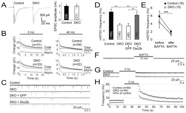
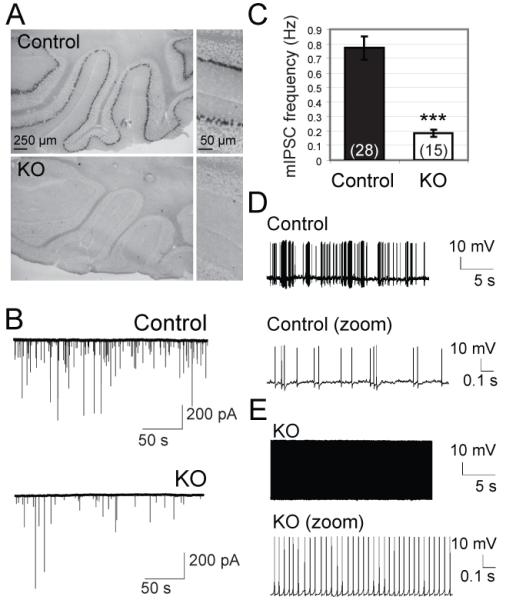
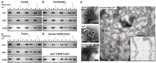
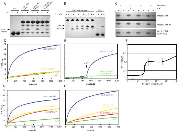
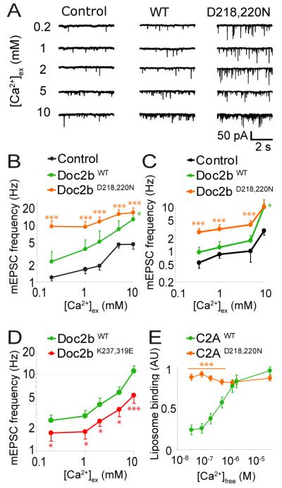
Synaptic vesicle fusion in brain synapses occurs in phases that are either tightly coupled to action potentials (synchronous), immediately following action potentials (asynchronous), or as stochastic events in the absence of action potentials (spontaneous). Synaptotagmin-1, -2, and -9 are vesicle-associated Ca2+ sensors for synchronous release. Here we found that double C2 domain (Doc2) proteins act as Ca2+ sensors to trigger spontaneous release. Although Doc2 proteins are cytosolic, they function analogously to synaptotagmin-1 but with a higher Ca2+ sensitivity. Doc2 proteins bound to N-ethylmaleimide-sensitive factor attachment receptor (SNARE) complexes in competition with synaptotagmin-1. Thus, different classes of multiple C2 domain-containing molecules trigger synchronous versus spontaneous fusion, which suggests a general mechanism for synaptic vesicle fusion triggered by the combined actions of SNAREs and multiple C2 domain-containing proteins.


Figures

Fig. 1
Fig. 1
Impaired spontaneous neurotransmission in Doc2a/b-deficient mice. Hippocampal neurons were cultured on microislands to promote self-innervation. (A) Averaged evoked excitatory postsynaptic currents (EPSCs) in control and DKO neurons. (B) Evoked EPSC charge (mean ± sem) during repeated stimulation at 5 or 40 Hz. The synchronous and asynchronous component were estimated as previously reported (24). (C) Representative traces of spontaneous EPSCs in wild-type or DKO cells. To rescue the phenotype, GFP or Doc2b were acutely expressed in DKO cells. (D) Average spontaneous release frequency in control and DKO cells and rescue by Doc2b overexpression. Cell numbers are indicated between brackets. **, P<0.01 (E) Average mEPSC frequency before and after intracellular loading of the Ca2+ chelator BAPTA. The average spontaneous release rate varied between experiments, but our experimental design prevents confounding effects thereof (fig. S2E) (F) Example trace representative for the data in (E), taken from a DKO cell before and after BAPTA loading. (G) Typical recordings before and after repetitive stimulation at 5 Hz. (H) Individual release events were binned in 1 s time intervals to monitor their average frequency immediately after repetitive stimulation. Squares were calculated as 50% of the frequency in control cells.
Fig. 2
Fig. 2
Reduced frequency of spontaneous events in Purkinje cells lacking Doc2b. (A) Doc2b mRNA was detected by in situ hybridization in cerebellar Purkinje cells from wildtype, but not Doc2b−/− (KO) mice. (B) Typical Voltage-clamp recordings in acute slices (C) Mean frequency of spontaneous inhibitory postsynaptic currents (mIPSCs) in KO mice and age-matched control littermates (n=15 and 28 cells; N=2 and 3 mice respectively). ***, p<0.0001. (D-E) Current clamp recordings of Purkinje cell firing patterns in controls (D, 8 cells from 3 mice) and KO cells (E, 7 cells from 2 mice).
Fig. 3
Fig. 3
Membrane-binding and curvature induction by Doc2b. (A-D) Liposome cosedimentation assays to characterize the membrane binding properties of the Doc2b C2A, C2B, C2AB, Doc2a C2AB and synaptotagmin-1 (Syt1) C2AB domains. Liposomes composed of the indicated lipids were incubated with the Doc2b C2A, C2B or C2AB domains. Liposome-bound protein was co-sedimented by ultracentrifugation. 8% of the supernatant and pellet fraction were run on 4-12% gradient gels. Proteins were visualized by Coomassie staining. S indicates unbound protein in the supernatant while P indicates liposome-bound and thus co-pelleted protein. No or very little protein sedimented in the absence of liposomes. (E) Ca2+- dependent tubulation of liposomes by the Doc2b C2AB domain. Folch liposomes were incubated in the absence or presence of Doc2b C2AB and/or Ca2+ and processed for electron microscopy by negative stain. The arrows indicate bundles of closely aligned tubules. Scale bar 100 nm. All data shown are representatives of at least 3 independent experiments. (PS: phosphatidylserine, PC: phosphatidylcholine, PIP2: phosphatidylinositol(4,5)bisphosphate)
Fig. 4
Fig. 4
SNARE complex binding and promotion of SNARE-dependent membrane fusion by Doc2b. (A) Coomassie stained gels showing a pull down experiment in the presence or absence of Ca2+ using the indicated Doc2b fusion proteins as bait and the purified SNARE complex as prey. The co-pelleted SNARE complex is indicated by an arrow. (B) Coomassie stained gels showing a pull down in the presence of Ca2+ using the GST-SNARE complex as bait and the Doc2b and synaptotagmin-1 C2AB domains as prey. GST served as control for unspecific binding by the C2AB domains of Doc2B and synaptotagmin-1, respectively. The co-pelleted Doc2B and synaptotagmin-1 C2AB domains are indicated by arrows. (C) Liposome co-sedimentation assay in the presence or absence of Ca2+ using the indicated proteins and liposomes composed of 20% PS, 70% PC and 10% cholesterol. (D) In vitro membrane fusion assay using reconstituted full length SNAREs in the absence or presence of Ca2+ and/or 7.5 μM Doc2b C2AB. (E) In vitro membrane fusion assay as in (D) but in the presence or absence of the soluble SNARE domain of synaptobrevin. Ca2+ was added at 500 s. (F) Ca2+ dose-dependence curve showing the change of fluorescence at 120 s in the reconstituted fusion assay in the presence of 7.5 μM Doc2b. Doc2b efficiently promotes fusion at sub-μM Ca2+ concentrations. The graph summarizes data from 3 independent experiments. (G) Comparison of the fusion promoting activities of the Doc2b, Doc2a and synaptotagmin-1 C2AB domains. (H) Fusion experiment using the indicated Doc2b C2AB proteins reveals that Ca2+-dependent membrane and SNARE-binding are required for efficient fusion promotion. All data shown are representatives of at least 3 independent experiments.
Fig. 5
Fig. 5
The Ca2+-dependence of Doc2b determines that of spontaneous release in rescued DKO neurons. (A) Typical mEPSC recordings in DKO cells rescued with GFP (control), Doc2bWT or Doc2bD218,220N. Spontaneous mEPSCs were recorded in network cultures of hippocampal neurons in the presence of TTX and gabazine. Increasing concentrations of extracellular Ca2+ were administered to the same cell. (B) Mean mEPSC frequencies ± sem in DKO cells expressing Doc2bWT (n=10), Doc2bD218,220N (n=26) or GFP as a control (n=21) (C) Mean mEPSC frequencies of the same constructs expressed in wildtype cells. The average mEPSC frequency varied between experiments, but our experimental design prevents confounding effects thereof (see Fig S2E) (D) Mean mEPSC frequencies in DKO cells expressing Doc2bWT (n=35) or mutant Doc2bK237,319E (n=31) (E) Ca2+-dependent liposome-binding by the isolated C2A domain of Doc2bWT and Doc2bD218,220N (n=6). *** (p<0.005); ** (p<0.01), * (p<0.05).

Similar articles

Cited by

References

    1. Geppert M, et al. Cell. 1994;79:717. - PubMed
    1. Fernandez-Chacon R, et al. Nature. 2001;410:41. - PubMed
    1. Martens S, Kozlov MM, McMahon HT. Science. 2007;316:1205. - PubMed
    1. Lynch KL, et al. Mol Biol Cell. 2008
    1. Pang ZP, Shin OH, Meyer AC, Rosenmund C, Sudhof TC. J Neurosci. 2006;26:12556. - PMC - PubMed

Publication types

MeSH terms