. 2010 Mar 2;107(9):4170-5.
doi: 10.1073/pnas.0914094107. Epub 2010 Feb 11.

Structure of sterol aliphatic chains affects yeast cell shape and cell fusion during mating

Affiliations

Structure of sterol aliphatic chains affects yeast cell shape and cell fusion during mating

Pablo S Aguilar et al. Proc Natl Acad Sci U S A. .

Abstract

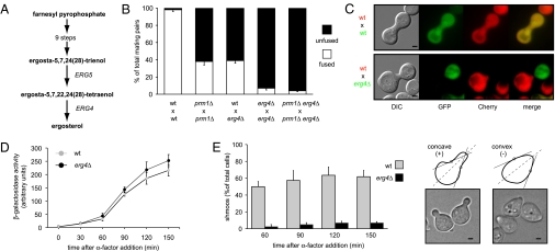
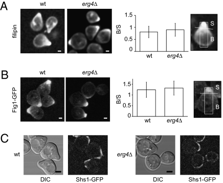
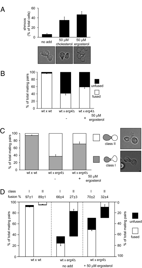
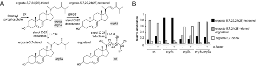
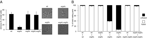
Under mating conditions, yeast cells adopt a characteristic pear-shaped morphology, called a "shmoo," as they project a cell extension toward their mating partners. Mating partners make contact at their shmoo tips, dissolve the intervening cell wall, and fuse their plasma membranes. We identified mutations in ERG4, encoding the enzyme that catalyzes the last step of ergosterol biosynthesis, that impair both shmoo formation and cell fusion. Upon pheromone treatment, erg4Delta mutants polarized growth, lipids, and proteins involved in mating but did not form properly shaped shmoos and fused with low efficiency. Supplementation with ergosterol partially suppressed the shmooing defect but not the cell fusion defect. By contrast, removal of the Erg4 substrate ergosta-5,7,22,24(28)-tetraenol, which accumulates in erg4Delta mutant cells and contains an extra double bond in the aliphatic chain of the sterol, restored both shmooing and cell fusion to wild-type levels. Thus, a two-atom change in the aliphatic moiety of ergosterol is sufficient to obstruct cell shape remodeling and cell fusion.


Conflict of interest statement

The authors declare no conflict of interest.

Figures

Fig. 1.
Fig. 1.
ERG4 mutant is defective in cell fusion. (A) Schematic representation of the ergosterol biosynthetic pathway highlighting the last two steps. (B) Quantitative cell fusion assays. All phenotypes were tested for both mating types, and the results were indistinguishable. Error bars indicate standard errors. (C) erg4Δ cells form morphologically abnormal mating pairs. MATα cells carrying cytoplasmic GFP were mixed with MATa cells carrying Pgk1-mCherry on nitrocellulose filters and incubated on YPD plates for 3 h at 30 °C. Fixed mating mixtures were then imaged by DIC and wide-field fluorescence microscopy. (Scale bars: 2 μm.) (D) erg4Δ cells preserve a normal pheromone response pathway. Strains harboring a FUS1-LACZ transcriptional reporter were grown and treated with 6 μM α-factor. At different time points, cells were harvested and assayed for β-galactosidase activity. Error bars indicate SEs. (E) erg4Δ cells are defective for shmoo formation. MATa cells were treated with α-factor, and aliquots were collected, fixed, and observed by bright-field microscopy at the indicated time points. For quantification (Left), cells that showed two convex edges along the axis of polarization (dotted lines) were scored as positive hits (Right). (Scale bars: 1 μm.)
Fig. 2.
Fig. 2.
erg4Δ cells show normal polarization of sterols and proteins. (A) Wild-type and erg4Δ MATa strains were treated with α-factor for 3 h, stained with filipin, and imaged by confocal microscopy (Left). (Scale bars: 1 μm.) Wild-type and erg4Δ MATa strains harboring either Fig1-GFP (B) or Shs1-GFP (C) were treated with α-factor for 3 h and then imaged by confocal microscopy. For A and B, fluorescence distribution along the longitudinal axis of cells was quantified as the fluorescence intensity ratio between the shmoo (S) and body (B) areas as described in Materials and Methods. Error bars indicate SEs. (Scale bars: B, 1 μm; C, 2 μm.)
Fig. 3.
Fig. 3.
Sterol supplementation partially suppresses erg4Δ shmooing and cell fusion defects. (A) erg4Δ cells were grown in anaerobic conditions in the presence of ergosterol and cholesterol. Cells were then harvested and treated with α-factor for 3 h, fixed, and observed under the microscope for shmoo formation quantification. Error bars indicate SEs. (BD) Cell fusion assay carried out with erg4Δ cells grown anaerobically either in the presence or absence of ergosterol and then mated with wild-type MATα cells bearing cytoplasmic GFP. Total cell fusion scores are shown in B. Different classes of mating pairs were classified depending on either the presence (Class I) or absence (Class II) of proper shmoo morphology in the erg4Δ partner, as shown in C. A breakdown of cell fusion scores for each class of mating pair, genotype, and growing conditions is shown in D. Error bars indicate standard errors.
Fig. 4.
Fig. 4.
Accumulation of different sterols in erg mutants. (A) Structures of the expected predominant sterols for each mutant strain. In the double mutant erg4Δ erg5Δ, the remaining sterol biosynthetic route ends in the production of ergosta-5,7,24(28)-trienol, whereas in the single mutant erg4Δ, the lack of sterol C-24 reductase activity produces the accumulation of ergosta-5,7,22,24(28)-tetraenol. As Erg4 sterol C-24 reductase activity can use ergosta-5,7,24(28)-trienol as a substrate, the predominant sterol in the erg5Δ strain should be ergosta-5,7-dienol. (B) Semiquantitative analysis of accumulated sterols. Free sterols were further purified and analyzed by mass spectrometry. The normalized peak areas of the protonated sterols in the MS1 are shown. Original spectra are depicted in Figs. S3S5.
Fig. 5.
Fig. 5.
erg5Δ suppresses erg4Δ. (A) Cells growing on YPD were treated with α-factor for 3 h, fixed, and examined under the microscope for quantification of shmooing efficiency. Error bars indicate standard errors. Bright-field microscopy representative images are shown at Right. (Scale bars: 1 μm.) (B) Quantitative cell fusion assays were performed testing both mating types for all mutants, and the results were indistinguishable. Error bars indicate SEs.

Similar articles

Cited by

References

    1. Chen EH, Grote E, Mohler W, Vignery A. Cell-cell fusion. FEBS Lett. 2007;581:2181–2193. - PubMed
    1. Ydenberg CA, Rose MD. Yeast mating: a model system for studying cell and nuclear fusion. Methods Mol Biol. 2008;475:3–20. - PubMed
    1. Herskowitz I. MAP kinase pathways in yeast: for mating and more. Cell. 1995;80:187–197. - PubMed
    1. Gammie AE, Brizzio V, Rose MD. Distinct morphological phenotypes of cell fusion mutants. Mol Biol Cell. 1998;9:1395–1410. - PMC - PubMed
    1. Philips J, Herskowitz I. Osmotic balance regulates cell fusion during mating in Saccharomyces cerevisiae. J Cell Biol. 1997;138:961–974. - PMC - PubMed

Publication types

LinkOut - more resources