. 2010 Mar;21(3):507-19.
doi: 10.1681/ASN.2008121259. Epub 2010 Feb 11.

Autoimmunity in membranous nephropathy targets aldose reductase and SOD2

Affiliations

Autoimmunity in membranous nephropathy targets aldose reductase and SOD2

Marco Prunotto et al. J Am Soc Nephrol. 2010 Mar.

Abstract

Glomerular targets of autoimmunity in human membranous nephropathy are poorly understood. Here, we used a combined proteomic approach to identify specific antibodies against podocyte proteins in both serum and glomeruli of patients with membranous nephropathy (MN). We detected specific anti-aldose reductase (AR) and anti-manganese superoxide dismutase (SOD2) IgG(4) in sera of patients with MN. We also eluted high titers of anti-AR and anti-SOD2 IgG(4) from microdissected glomeruli of three biopsies of MN kidneys but not from biopsies of other glomerulonephritides characterized by IgG deposition (five lupus nephritis and two membranoproliferative glomerulonephritis). We identified both antigens in MN biopsies but not in other renal pathologies or normal kidney. Confocal and immunoelectron microscopy (IEM) showed co-localization of anti-AR and anti-SOD2 with IgG(4) and C5b-9 in electron-dense podocyte immune deposits. Preliminary in vitro experiments showed an increase of SOD2 expression on podocyte plasma membrane after treatment with hydrogen peroxide. In conclusion, our data support AR and SOD2 as renal antigens of human MN and suggest that oxidative stress may drive glomerular SOD2 expression.


Figures

Figure 1.
Figure 1.
Sera of MN patients show presence of antibodies against podocyte proteins. (A through C) 2D electrophoresis analysis of podocyte membrane extracts and Western blot analysis of MN serum for anti-podocyte antibodies. (A) Representative 2D map of podocyte membrane extracts stained by colloidal Coomassie. Numbers correspond to identified proteins as displayed in Table 2; spots 1, 2, 3, and 4 correspond to SOD2, and spots 13, 14, and 15 correspond to AR. (B and C) Western blot analysis of MN serum for anti-podocyte antibodies. Sera of patients with MN were largely positive for SOD2 (B and C); only a few were positive for AR (C). Representative normal sera (n = 10) run in parallel to MN samples were negative and have been reported as control. (D through I) Expression of AR and SOD2 on cultured podocytes. Nonpermeabilized human podocyte cell line stained for AR (D) or SOD2 (G) and MN sera (10% in medium) incubated with cells (E and H). (F and I) Merged images. Arrows in merged images indicate the coexpression of AR or SOD2 with MN IgG. Magnification, ×630.
Figure 2.
Figure 2.
Circulating anti-AR and anti-SOD2 antibodies are present in the sera of MN patients and completely absent in FSGS. (A and C) MN sera (n = 24, first two rows) screened for the presence of autoantibodies were positive for both anti-AR (A) and anti-SOD2 (C) IgG4. Intensity was mild or absent in normal sera (n = 24, bottom rows) and completely absent in FSGS (NS). St., calibration curve. (B and D) Corresponding dot graphs plotting OD for both anti-AR (B) and anti-SOD2 (D) IgG4. Nonparametric statistical tests (Mann-Whitney U test) showed a statistical difference between MN and normal sera (N) in all cases (P < 0.01 for anti-AR and P < 0.001 for anti-SOD IgG4; the difference between MN and FSGS was P < 0.001 in all cases).
Figure 3.
Figure 3.
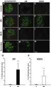
AR and SOD2 are expressed in renal biopsies of MN patients. (A through N) Representative images of renal biopsy specimens from normal patients (A through C), and patients with MN (D through F), MCD (G through I), and MN secondary to neoplasia (L through N) stained for synaptopodin (A, D, G, and L), AR (B, E, H, and M), or SOD2 (C, F, I, and N). AR and SOD2 staining is absent in tissues from normal individuals normal and from patients with MCD and only very weakly expressed in tissues from patients with MN secondary to neoplasia, whereas a marked glomerular staining for both proteins is evident in patients with MN. Synaptopodin is included to highlight glomerular structures. Bar = 200 μ. (O and P) Morphometric results of glomerular expression of AR and SOD2 in patients with MN. Bar graph shows glomerular expression of AR (■) and SOD2 (□), as measured by morphometric analysis on tissues from studied cases. Results are expressed as percentage of the positive area for each considered antibody on total glomerular area. The protein expression for both was increased in patients with MN, with high statistical significance (P < 0.001) when compared with control patients with a higher expression of AR in all considered glomeruli. Magnification, ×400.
Figure 4.
Figure 4.
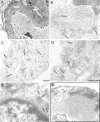
AR is present in GBM and in subepithelial immune deposits. (A and B) AR is present in GBM as shown by several scattered gold particles in the context of electron-dense subepithelial immune deposits (arrows). (C through E) AR was also observed in the cytoplasm and the plasma membrane of podocyte bodies (Po). (E) AR is not expressed at the nuclear (N) level; instead, AR is present in the cytoplasm as indicated by arrows. (F) Blank sample lacking primary antibody was totally negative (see also Supplemental Figure 5, A, C, and E). EN, endothelium. Magnifications: ×8900 in A; ×56,000 in B, D, and E; ×36,000 in C; ×44,000 in F.
Figure 5.
Figure 5.
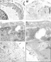
SOD2 is present in GBM and on podocyte plasma membrane. (A and B) SOD2 was detected in electron-dense immune deposits within the GBM (arrows) as shown by several scattered gold particles. (C and D) SOD2 was also observed in the cytoplasm and on plasma membrane of podocyte bodies (Po). (F) Blank sample omitting specific primary antibody was totally negative (see also Supplemental Figure 5, B, D, and F), EN, endothelium; n, podocyte nucleus. Magnifications: ×8900 in A; ×44,000 in B and C; ×71,000 in D; ×51,000 in E; ×36,000 F.
Figure 6.
Figure 6.
AR and SOD2 co-localize with IgG4 in glomeruli of MN patient biopsies. (A, B, D, and E) Confocal images of a renal biopsy specimen from a patient with MN. Double IF staining was evaluated for AR (A), SOD2 (D) and IgG4 (B and E). Merged images are reported in C and F and in p1 and p2. Both AR and SOD2 were co-localized with IgG4 along the capillary walls. Magnification, ×630.
Figure 7.
Figure 7.
AR and SOD2 co-localize with C5b-9 in glomeruli of MN patient biopsies. Confocal images of a renal biopsy specimen from MN. (A, B, D, and E) Double IF staining for AR (A), SOD2 (D), and C5b-9 (B and E). Merged images are reported in C and F and in p1 and p2. Both AR and SOD2 co-localize in immune deposits. Magnification, ×630.
Figure 8.
Figure 8.
Glomerular eluates from MN patients contain antibodies against podocyte antigens. Podocyte extracts were separated by electrophoresis in reducing (a through e) and nonreducing (f through h) conditions and were studied for reactivity with eluates from microdissected glomeruli. Thirty micrograms of protein was used for the analysis: Part was analyzed with silver stain (a and f), and part was incubated with glomerular eluates from patients with MN and then developed with anti-IgG4 (b and g). Eight antigens were recognized in reducing conditions and characterized by mass spectrometry as AR, SOD, and vimentin. Three more bands with a molecular weight of 200, 170, and 155 kD were detected in nonreducing conditions. (c and h) Control lines for Western blot in which glomerular eluates were omitted. (d) Western blot with anti-AR antibody. (e) Western blot with SOD2 antibodies.
Figure 9.
Figure 9.
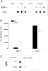
Glomerular eluates from MN patients contain antibodies against AR and SOD2. (A) Glomeruli showed presence of specific IgG4 antibodies against AR and SOD2. IgGs were also present at a lower titer. Controls were incubated with human serum albumin (HSA) instead of glomerular eluates. (B) Corresponding bar graph shows dot-spot OD (in arbitrary units) for anti-AR (■) and anti-SOD2 (□) specific IgG4. (C) Competition experiment using the same glomerular eluates as in A and increasing amount of r-AR and r-SOD2 from 5 to 15 ng.
Figure 10.
Figure 10.
Glomerular eluates from other pathologies do not contain antibodies against AR or SOD2. Dot blot of eluates from microdissected glomeruli of a normal kidney (N) and patients (n = 7) with other glomerulonephritis implying IgG deposition in glomeruli: Systemic lupus nephritis (SLE; n = 5) and membranoproliferative glomerulonephritis (MPGN; n = 2). For comparison, two patients with MN were displayed. Anti-AR and anti-SOD IgG4 was determined as described in the Concise Methods section. Total IgG titer was determined with dot blot to nonsaturated nitrocellulose membrane, and proteins were revealed with biotinylated anti-human IgG.
Figure 11.
Figure 11.
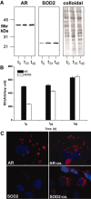
In vitro oxidation modifies AR and SOD2 expression on plasma membrane. (A through C) Western blot (A), corresponding bar graph (B; ■, AR; □, SOD2) and IF staining (C) for AR or SOD2 upon H2O2 treatment. H2O2 treatment increased SOD2 expression at 24 and 48 hours, whereas AR was only slightly affected. IF on nonpermeabilized podocytes confirmed Western blot results. Magnification, ×630.

Similar articles

Cited by

References

    1. Wasserstein AG: Membranous glomerulonephritis. J Am Soc Nephrol 8: 664–674, 1997 - PubMed
    1. Kerjaschki D: Pathomechanisms and molecular basis of membranous glomerulopathy. Lancet 364: 1194–1196, 2004 - PubMed
    1. Neale TJ, Ojha PP, Exner M, Poczewski H, Ruger B, Witztum JL, Davis P, Kerjaschki D: Proteinuria in passive Heymann nephritis is associated with lipid peroxidation and formation of adducts on type IV collagen. J Clin Invest 94: 1577–1584, 1994 - PMC - PubMed
    1. Neale TJ, Ullrich R, Ojha P, Poczewski H, Verhoeven AJ, Kerjaschki D: Reactive oxygen species and neutrophil respiratory burst cytochrome b558 are produced by kidney glomerular cells in passive Heymann nephritis. Proc Natl Acad Sci U S A 90: 3645–3649, 1993 - PMC - PubMed
    1. Rastaldi MP, Candiano G, Musante L, Bruschi M, Armelloni S, Rimoldi L, Tardanico R, Sanna-Cherchi S, Ferrario F, Montinaro V, Haupt R, Parodi S, Carnevali ML, Allegri L, Camussi G, Gesualdo L, Scolari F, Ghiggeri GM: Glomerular clusterin is associated with PKC-alpha/beta regulation and good outcome of membranous glomerulonephritis in humans. Kidney Int 70: 477–485, 2006 - PubMed

Publication types

MeSH terms