. 2010 Apr;30(4):787-95.
doi: 10.1161/ATVBAHA.109.202051. Epub 2010 Feb 11.

Macrophage LRP-1 controls plaque cellularity by regulating efferocytosis and Akt activation

Affiliations

Macrophage LRP-1 controls plaque cellularity by regulating efferocytosis and Akt activation

Patricia G Yancey et al. Arterioscler Thromb Vasc Biol. 2010 Apr.

Abstract

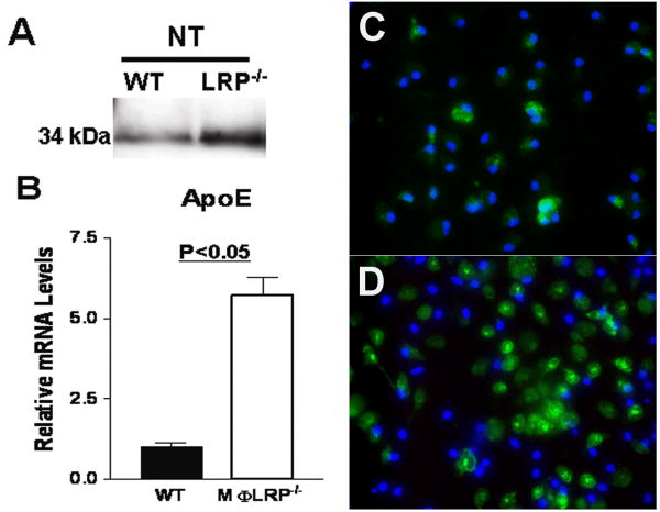
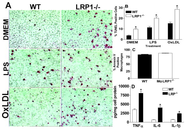
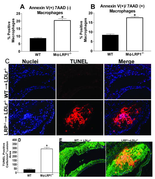
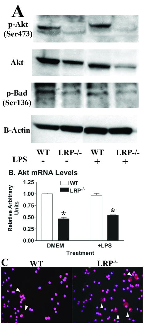
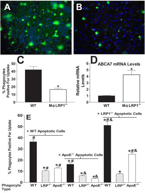
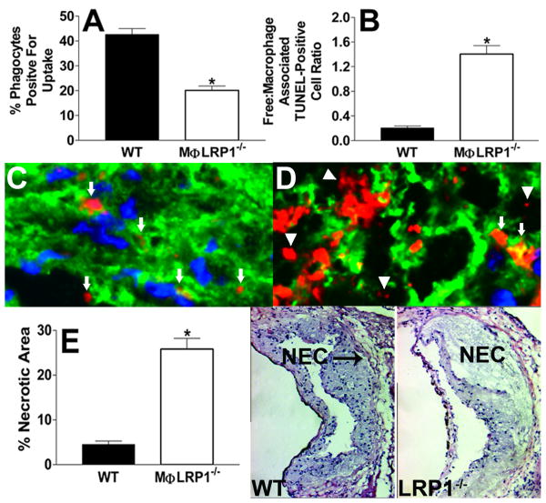
Objective: The balance between apoptosis susceptibility and efferocytosis of macrophages is central to plaque remodeling and inflammation. LRP-1 and its ligand, apolipoprotein E, have been implicated in efferocytosis and apoptosis in some cell types. We investigated the involvement of the macrophage LRP-1/apolipoprotein E axis in controlling plaque apoptosis and efferocytosis. Method and Results- LRP-1(-/-) macrophages displayed nearly 2-fold more TUNEL positivity compared to wild-type cells in the presence of DMEM alone or with either lipopolysaccharide or oxidized low-density lipoprotein. The survival kinase, phosphorylated Akt, was barely detectable in LRP-1(-/-) cells, causing decreased phosphorylated Bad and increased cleaved caspase-3. Regardless of the apoptotic stimulation and degree of cell death, LRP-1(-/-) macrophages displayed enhanced inflammation with increased IL-1 beta, IL-6, and tumor necrosis factor-alpha expression. Efferocytosis of apoptotic macrophages was reduced by 60% in LRP-1(-/-) vs wild-type macrophages despite increased apolipoprotein E expression by both LRP-1(-/-) phagocytes and wild-type apoptotic cells. Compared to wild-type macrophage lesions, LRP-1(-/-) lesions had 5.7-fold more necrotic core with more dead cells not associated with macrophages.

Conclusions: Macrophage LRP-1 deficiency increases cell death and inflammation by impairing phosphorylated Akt activation and efferocytosis. Increased apolipoprotein E expression in LRP-1(-/-) macrophages suggests that the LRP-1/apolipoprotein E axis regulates the balance between apoptosis and efferocytosis, thereby preventing necrotic core formation.


Conflict of interest statement

Disclosures: Sergio Fazio (≥$10 000), Patricia G. Yancey (≥$10 000), and MacRae F. Linton (≥$10 000) received research grant RO1 HL57986.

Figures

Figure 1
Figure 1
Detection of apoE protein in the medium (A), apoE mRNA levels in cells (B), and immunohistochemistry for apoE (green) and nuclei (DAPI, blue) in WT (C) or LRP1-/- (D) macrophages after 24h in serum-free DMEM. *p<0.05, Student's t test.
Figure 2
Figure 2
TUNEL positive macrophages (A and B) after 24h in DMEM alone or with LPS (50ng/ml) or oxidized LDL (50μg/ml). Annexin V positive cells (C) and medium cytokine levels (D) after 24h in DMEM with staurosporine (50μg/ml). *p<0.05 between the two groups, Student's t test (A-D). Data represent 2 (C-D) or 3 (A-B) experiments.
Figure 3
Figure 3
Peritoneal macrophages positive for annexinV alone(A) or annexinV and 7AAD (B). TUNEL analyses of aortic sections from WT (n=5) and MΦLRP1-/- (n=4) BM recipient LDLr-/- mice fed western diet for 16 weeks (C). Micrographs show nuclei (DAPI, blue), TUNEL positive staining (red) and merged images. Quantitation of TUNEL results (D) and co-localization of macrophage stain (green) in TUNEL-positive regions of arteries from mice with LRP1-/- BM (E). *p<0.05, Mann-Whitney test
Figure 4
Figure 4
(A) Western blot of cell pAkt(serine 473), total Akt, pBad(serine 136), and β-actin and (B) Akt1 mRNA levels in WT or LRP1-/- macrophages after 24h in DMEM alone or with LPS (50ng/ml). (C) Immunohistochemical detection of activated caspase-3 (red) after 24h in DMEM. DAPI (pink) staining identifies nuclei. *p<0.05, Student's t test (B) and all data represent 2 experiments.
Figure 5
Figure 5
Efferocytosis of apoptotic (CFDA-SE/green label) WT cells by WT (A) or LRP1-/- (B) macrophages. DAPI (blue) staining identifies nuclei. (C) CFDA-SE positive phagocytes as percentage of total macrophages. (D) ABCA7 mRNA after 24h in DMEM. *p<0.05, Student's t test. (E) Phagocytosis of CFDA-SE labeled WT, apoE-/-, or LRP1-/- apoptotic macrophages by WT, apoE-/-, or LRP1-/- efferocytes. Differences (p<0.05) were marked as follows: * versus the WT/WT condition; # versus the apoE-/-/apoE-/- condition; & versus the apoE-/-/WT condition. ANOVA with Bonferroni's post test. Data represent two (D, E) or four (C) experiments.
Figure 6
Figure 6
Percent of macrophages positive for efferocytosis of apoptotic CFDA-SE/labeled WT cells (A) 1h post injection into the peritoneal cavity of WT (n=3) or MΦLRP1-/- (n=4) mice as determined by flow cytometry. Quantitation of the ratio of free versus macrophage associated TUNEL positive cells (B) in aortic sections from WT (n=5) and MΦLRP1-/- (n=4) BM recipient LDLr-/- mice fed western diet for 16 weeks. (C, D) TUNEL positive cells in aortic sections from WT (C) or MΦLRP1-/- (D) BM recipient LDLr-/- mice. Cells are either free (triangles) or associated with viable macrophages (arrows). (E) Quantitation of necrotic area in aortic sections from WT (n=5) and MΦLRP1-/- (n=4) BM recipient LDLr-/- mice. Images show hematoxylin and eosin staining of aortic root sections. NEC denotes necrotic area (E). *p<0.05, Mann Whitney test.

Similar articles

Cited by

References

    1. Herz J, Clouthier DE, Hammer RE. LDL receptor-related protein internalizes and degrades uPA-PAI-1 complexes and is essential for embryo implantation. Cell. 1992;71:411–421. - PubMed
    1. Rohlmann A, Gotthardt M, Hammer RE, Herz J. Inducible inactivation of hepatic LRP gene by cre-mediated recombination confirms role of LRP in clearance of chylomicron remnants. Journal of Clinical Investigation. 1998;101:689–695. - PMC - PubMed
    1. Overton CD, Yancey PG, Major AS, Linton MF, Fazio S. Deletion of Macrophage LDL Receptor-Related Protein Increases Atherogenesis in the Mouse. Circ Res. 2007;100:670–677. - PubMed
    1. Li Y, Gerbod-Giannone MC, Seitz H, Cui D, Thorp E, Tall AR, Matsushima GK, Tabas I. Cholesterol-induced apoptotic macrophages elicit an inflammatory response in phagocytes, which is partially attenuated by the Mer receptor. J Biol Chem. 2006;281:6707–6717. - PubMed
    1. Boullier A, Li Y, Quehenberger O, Palinski W, Tabas I, Witztum JL, Miller YI. Minimally oxidized LDL offsets the apoptotic effects of extensively oxidized LDL and free cholesterol in macrophages. Arterioscler Thromb Vasc Biol. 2006;26:1169–1176. - PubMed

Publication types

MeSH terms