. 2010 Mar 17;29(6):1091-104.
doi: 10.1038/emboj.2010.7. Epub 2010 Feb 11.

Genetic analysis of Ras signalling pathways in cell proliferation, migration and survival

Affiliations

Genetic analysis of Ras signalling pathways in cell proliferation, migration and survival

Matthias Drosten et al. EMBO J. .

Abstract

We have used mouse embryonic fibroblasts (MEFs) devoid of Ras proteins to illustrate that they are essential for proliferation and migration, but not for survival, at least in these cells. These properties are unique to the Ras subfamily of proteins because ectopic expression of other Ras-like small GTPases, even when constitutively active, could not compensate for the absence of Ras proteins. Only constitutive activation of components of the Raf/Mek/Erk pathway was sufficient to sustain normal proliferation and migration of MEFs devoid of Ras proteins. Activation of the phosphatidylinositol 3-kinase (PI3K)/PTEN/Akt and Ral guanine exchange factor (RalGEF)/Ral pathways, either alone or in combination, failed to induce proliferation or migration of Rasless cells, although they cooperated with Raf/Mek/Erk signalling to reproduce the full response mediated by Ras signalling. In contrast to current hypotheses, Ras signalling did not induce proliferation by inducing expression of D-type Cyclins. Rasless MEFs had normal levels of Cyclin D1/Cdk4 and Cyclin E/Cdk2. However, these complexes were inactive. Inactivation of the pocket proteins or knock down of pRb relieved MEFs from their dependence on Ras signalling to proliferate.


Conflict of interest statement

The authors declare that they have no conflict of interest.

Figures
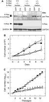
Figure 1
Figure 1
Proliferative properties of MEFs expressing single Ras loci. (A) Western blot analysis of H-Ras−/−;N-Ras−/−;K-Raslox/lox;RERTert/ert, H-Ras+/+;N-Ras−/−;K-Raslox/lox;RERTert/ert, and H-Ras−/−;N-Ras+/+;K-Raslox/lox;RERTert/ert MEFs 3 days after infection with Adeno-GFP or Adeno-Cre. The pan-Ras antibody recognizes all Ras proteins. GAPDH expression was used as a loading control. (B) Growth curve of primary (passage 1) wild type (solid squares), H-Ras−/−;N-Ras−/−;K-Raslox/lox;RERTert/ert MEFs infected with Adeno-GFP (open squares) and H-Ras+/+;N-Ras−/−;K-Raslox/lox;RERTert/ert MEFs (open triangles) as well as H-Ras−/−;N-Ras+/+;K-Raslox/lox;RERTert/ert MEFs (open circles) infected with Adeno-Cre to remove the conditional K-Raslox alleles. Ras mutant MEFs were derived from two independent embryos. Error bars indicate standard deviation. (C) Immortalization of wild type (solid squares), H-Ras−/−;N-Ras−/−;K-Raslox/lox;RERTert/ert MEFs (open squares), H-Ras+/+;N-Ras−/−;K-Raslox/lox;RERTert/ert MEFs (open triangles) and H-Ras−/−;N-Ras+/+;K-Raslox/lox;RERTert/ert MEFs (open circles) following a 3T3 protocol. H-Ras+/+;N-Ras−/−;K-Raslox/lox;RERTert/ert and H-Ras−/−;N-Ras+/+;K-Raslox/lox;RERTert/ert MEFs were maintained in the presence of 4OHT to achieve excision of the K-Raslox alleles. Ras mutant MEFs were derived from two independent embryos. Error bars indicate standard deviation.
Figure 2
Figure 2
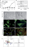
Ras proteins are essential for cell proliferation and migration. (A) Proliferation of immortal K-Raslox MEFs left untreated (open circles) or exposed to 4OHT for the indicated time to eliminate the conditional K-Raslox alleles (solid circles). Error bars indicate standard deviation. Inset depicts a Southern blot analysis showing excision of the conditional K-Raslox alleles (lox) in the presence of 4OHT (knock out, KO). (B) Western blot analysis showing Ras protein expression in K-Raslox cells either left untreated or exposed to 4OHT for 7 or 14 days. Wild-type (WT) MEFs were used as control. Migration of the indicated proteins is shown by arrowheads. Total Erk proteins were used as loading control. (C) Top: bright field microscopic images of K-Raslox MEFs left untreated (K-Raslox) and treated with 4OHT for 14 days (Rasless). Scale bar represents 100 μm. Middle: confocal microscopy images of microtubule organization in K-Raslox MEFs left untreated (K-Raslox) or 14 days after treatment with 4OHT (Rasless) as determined by α-tubulin staining (green) and DAPI counterstaining (blue). Scale bar represents 50 μm. Bottom: confocal microscopy images of actin stress fibres and focal adhesions in K-Raslox MEFs left untreated (K-Raslox) or exposed to 4OHT for 14 days (Rasless) as determined by Phalloidin (green) and Paxillin staining (red). DAPI counterstaining (blue) indicates nuclei. Scale bar represents 50 μm. (D) Migration tracks of individual K-Raslox MEFs left untreated (K-Raslox) (left) or 14 days after treatment with 4OHT (Rasless) (right) as determined by time-lapse microscopy. Migration was tracked for 14 h (n=8).
Figure 3
Figure 3
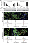
The proliferation and migration defects of Rasless MEFs are reversible. (A) Upper panel: percentages of cells in S phase as indicated by BrdU incorporation in (left) Tet-Off-K-Ras and (right) Tet-Off-K-RasG12V cells after addition and subsequent withdrawal of doxycyline at the indicated time points. Error bars indicate standard deviation. Lower panel: western blot analysis of K-Ras expression at the indicated times. GAPDH expression served as loading control. (B, C) Upper panels: migration defects and cytoskeletal alterations in (B) Tet-Off-K-Ras and (C) Tet-Off-K-RasG12V cells before addition of doxycycline (Day 0), 10 days after treatment with doxycycline (+Dox/Days 0–10) and four additional days after doxycyline withdrawal (−Dox/Days 10–14) as determined by Phalloidin (green) and Paxillin staining (red). DAPI counterstaining (blue) indicates nuclei. Scale bar represents 75 μm. Lower panels: migration tracks from individual (B) Tet-Off-K-Ras and (C) Tet-Off-K-RasG12V cells as determined by time-lapse microscopy. Migration was tracked for 14 h (n=8).
Figure 4
Figure 4
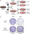
(A) Schematic representation of the assay used to identify genes capable of inducing cell proliferation in the absence of Ras proteins. (B) Representative plates from colony formation assays. K-Raslox MEFs were infected with empty retroviruses (control cultures) or retroviruses expressing a K-Ras4B cDNA (effector cultures). After antibiotic selection, cells were plated in the absence (−) or presence (+) of 4OHT and allowed to form colonies for 14 days. The tiny colonies observed in the control Rasless plate consist of small groups of cells (8–32 cells on average) resulting from limited cell proliferation until the K-Raslox alleles are completely excised. None of these cells was able to proliferate for extended periods of time nor on transfer to other plates. Cells were fixed with glutaraldehyde and stained with crystal violet.
Figure 5
Figure 5
Expression of constitutively active Ras-related small GTPases do not sustain proliferation of Rasless MEFs. (A) Colony-formation assay using K-Raslox MEFs infected with retroviruses expressing the indicated cDNAs. Error bars indicate standard deviation. (B) Western blot analysis showing levels of expression of the proteins encoded by the corresponding cDNAs indicated in (A). GAPDH expression served as a loading control. Migration of the indicated proteins is shown by arrowheads. (C) Western blot analysis of Mek and Akt phosphorylation in K-Raslox MEFs infected with retroviruses encoding the indicated Ras-related proteins incubated in the presence (+) or absence (−) of 4OHT. Indicated cultures were either maintained in the presence of 10% FBS (+) or subjected to serum withdrawal (0.1% FBS for 24 h) (−). Migration of the indicated proteins is shown by arrowheads. Total Mek1 and Akt proteins served as loading controls.
Figure 6
Figure 6
Constitutive activation of the Raf/Mek/Erk pathway drives proliferation of Rasless MEFs. (A) Colony-formation assay using K-Raslox MEFs infected with retroviruses expressing the indicated cDNAs. p16 shRNA indicates a retrovirus encoding an shRNA directed against p16INK4a-specific sequences. Error bars indicate standard deviation. (B) Western blot analysis showing levels of expression of the indicated H-Ras effector mutant proteins probed with a pan-Ras antibody. Expression of the corresponding H-Ras or K-Ras proteins is indicated by arrowheads. GAPDH expression served as a loading control. (C) Western blot analysis showing expression of the indicated H-RasG12V or RafCAAX proteins in K-Raslox MEFs co-infected with a retrovirus encoding an shRNA directed against p16INK4a-specific sequences (+). Expression of p16INK4a is also indicated. Cells were left untreated (−) or exposed to 4OHT (+). Probing antibodies are indicated in the right panel. Migration of the corresponding proteins is indicated by solid arrowheads. Open arrowheads indicate migration of the endogenous B-RafCAAX and C-RafCAAX proteins. GAPDH expression served as loading control. (D) Western blot analysis showing expression of H-RasG12V, Mek1Q56P and ERK2MEK1LA (EMLA) in K-Raslox MEFs left untreated (−) or exposed (+) to 4OHT. Probing antibodies are indicated in the right panel. Migration of the corresponding proteins is indicated by solid arrowheads. Open arrowheads indicate migration of endogenous Mek1 and Erk1/2 proteins. GAPDH expression served as loading control.
Figure 7
Figure 7
Constitutive activation of the RalGDS and PI3K pathways fails to confer proliferative properties to Rasless cells. (A) Colony-formation assay using K-Raslox MEFs infected with retroviruses expressing the indicated cDNAs or an shRNA directed against PTEN-specific sequences. Error bars indicate standard deviation. (B) Western blot analysis of K-Raslox MEFs infected with the indicated retroviruses. Cells were left untreated (−) or exposed (+) to 4OHT. Probing antibodies are listed in the right panel. Migration of the corresponding proteins is indicated by solid arrowheads. GAPDH expression served as loading control.
Figure 8
Figure 8
Role of cell cycle regulators on the proliferative properties of Rasless cells. (A) Expression levels of cell cycle Cyclins in K-Raslox MEFs exposed to 4OHT for the indicated times as determined by western blot analysis. Loss of K-Ras expression is illustrated in the upper panel. α-Tubulin expression served as loading control. (B) Identification of Cyclin D1/Cdk4 (upper panel) and Cyclin E1/Cdk2 (lower panel) complexes by immunoprecipitation with antibodies against Cyclin D1 (upper panel) and Cyclin E (lower panel) in K-Raslox and Rasless cells; 1/16th of the whole cell extract (WCE) used for immunoprecipitation of K-Raslox MEFs was loaded as a control. Additional controls included mock (M) immunoprecipitates obtained from K-Raslox MEFs by incubating without primary antibody and 1/16th of WCE from Cdk2−/−;Cdk4−/− MEFs (Cdk2/4dKO). (C) Analysis of pRb phosphorylation in K-Raslox and Rasless cells by western blot analysis using antibodies against PSer807/811-phosphorylated pRb. Total pRb was used as a loading control. (D) Colony-formation assay using K-Raslox MEFs infected with retroviruses expressing the indicated cDNAs or an shRNA directed against pRb-specific sequences. Error bars indicate standard deviation. (E) Upper panels: western blot analysis of Cyclin D1, Cyclin E1, Cyclin A2 and Cyclin B1 expression in K-Raslox MEFs infected with an empty vector or with retroviruses encoding wild-type Cyclin D1, Cyclin D1T286A, Cyclin E1, Cyclin A2 and Cyclin B1. Migration of the indicated proteins is shown by arrowheads. α-Tubulin (T) or GAPDH (G) expression served as loading controls. Lower panels: quantification of protein expression levels from at least two independent experiments.

References

    1. Aktas H, Cai H, Cooper GM (1997) Ras links growth factor signaling to the cell cycle machinery via regulation of cyclin D1 and the Cdk inhibitor p27KIP1. Mol Cell Biol 17: 3850–3857 - PMC - PubMed
    1. Bélanger LF, Roy S, Tremblay M, Brott B, Steff AM, Mourad W, Hugo P, Erikson R, Charron J (2003) Mek2 is dispensable for mouse growth and development. Mol Cell Biol 23: 4778–4787 - PMC - PubMed
    1. Bissonauth V, Roy S, Gravel M, Guillemette S, Charron J (2006) Requirement for Map2k1 (Mek1) in extra-embryonic ectoderm placentogenesis. Development 133: 3429–3440 - PubMed
    1. Bottorff D, Stang S, Agellon S, Stone JC (1995) RAS signaling is abnormal in a c-raf1 MEK1 double mutant. Mol Cell Biol 15: 5113–5122 - PMC - PubMed
    1. Buday L, Downward J (2008) Many faces of Ras activation. Biochim Biophys Acta 1786: 178–187 - PubMed

Publication types

MeSH terms

Substances