. 2010 Jan 15:3:2.
doi: 10.1186/1754-6834-3-2.

Furfural induces reactive oxygen species accumulation and cellular damage in Saccharomyces cerevisiae

Affiliations

Furfural induces reactive oxygen species accumulation and cellular damage in Saccharomyces cerevisiae

Sandra A Allen et al. Biotechnol Biofuels. .

Abstract

Background: Biofuels offer a viable alternative to petroleum-based fuel. However, current methods are not sufficient and the technology required in order to use lignocellulosic biomass as a fermentation substrate faces several challenges. One challenge is the need for a robust fermentative microorganism that can tolerate the inhibitors present during lignocellulosic fermentation. These inhibitors include the furan aldehyde, furfural, which is released as a byproduct of pentose dehydration during the weak acid pretreatment of lignocellulose. In order to survive in the presence of furfural, yeast cells need not only to reduce furfural to the less toxic furan methanol, but also to protect themselves and repair any damage caused by the furfural. Since furfural tolerance in yeast requires a functional pentose phosphate pathway (PPP), and the PPP is associated with reactive oxygen species (ROS) tolerance, we decided to investigate whether or not furfural induces ROS and its related cellular damage in yeast.

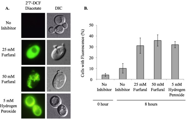
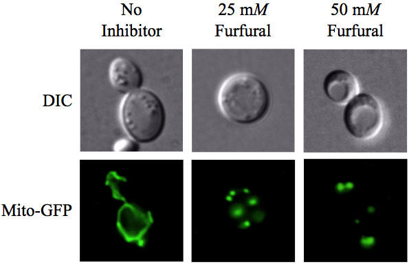
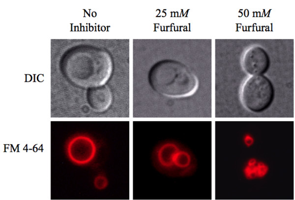
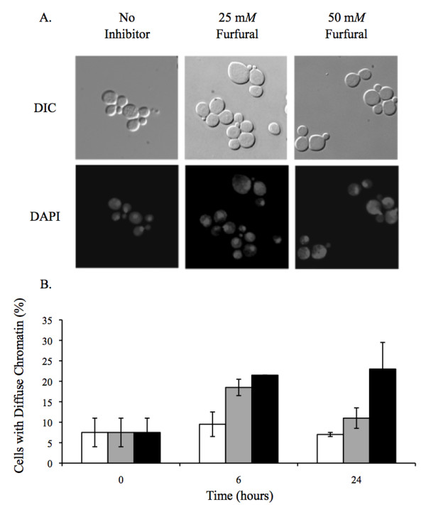
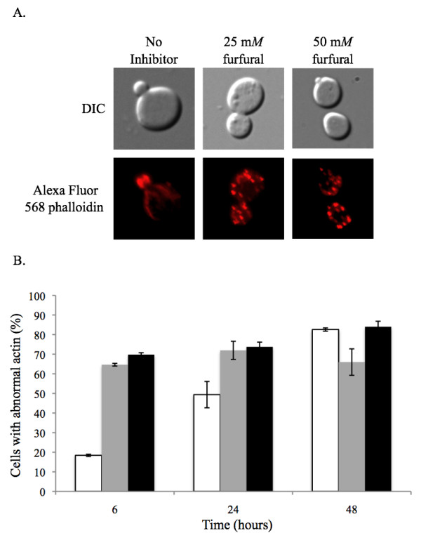
Results: We demonstrated that furfural induces the accumulation of ROS in Saccharomyces cerevisiae. In addition, furfural was shown to cause cellular damage that is consistent with ROS accumulation in cells which includes damage to mitochondria and vacuole membranes, the actin cytoskeleton and nuclear chromatin. The furfural-induced damage is less severe when yeast are grown in a furfural concentration (25 mM) that allows for eventual growth after an extended lag compared to a concentration of furfural (50 mM) that prevents growth.

Conclusion: These data suggest that when yeast cells encounter the inhibitor furfural, they not only need to reduce furfural into furan methanol but also to protect themselves from the cellular effects of furfural and repair any damage caused. The reduced cellular damage seen at 25 mM furfural compared to 50 mM furfural may be linked to the observation that at 25 mM furfural yeast were able to exit the furfural-induced lag phase and resume growth. Understanding the cellular effects of furfural will help direct future strain development to engineer strains capable of tolerating or remediating ROS and the effects of ROS.


Figures

Figure 1
Figure 1
Furfural induces the accumulation of reactive oxygen species (ROS). Exponentially growing yeast cells were treated with no inhibitor, 25 mM furfural, 50 mM furfural or 5 mM hydrogen peroxide (positive control for ROS). (A) Representative images of cells stained with the ROS indicator dye 2'7' DCF-diacetate (left column) and differential interference contrast (DIC) (right column) are shown. Images of cells were at the 8 h time point. (B) Percent of yeast cells that stained positive for ROS by 2'7' DCF-diacetate at 0 h and 8 h. Data represent an average of five experiments with standard error indicated. At each time point at least 100 cells were examined.
Figure 2
Figure 2
Furfural causes internal cellular damage. Exponentially growing yeast cells grown with either no inhibitor (1st row) or 25 mM furfural (2nd and 3rd rows) were fixed and thin-sectioned for transmission electron microscopy analysis after they had been exposed to furfural for 8 h. Mitochondria are indicated by arrows, vacuoles by asterisks, and nuclei by N.
Figure 3
Figure 3
Furfural causes mitochondrial membrane morphology to go from tubules to aggregates. Exponentially growing yeast cells expressing mitochondrial targeted green fluorescent protein were untreated or treated with 25 mM or 50 mM furfural. Representative images of yeast with no inhibitor (left column; tubular), exposed to 25 mM furfural (middle column; evenly distributed fragments) or 50 mM furfural (right column; aggregated) are shown. Images of cells were taken 6 h after furfural treatment.
Figure 4
Figure 4
Furfural causes vacuoles to go from large single organelles to several smaller ones. Exponentially growing yeast cells were either untreated or treated with 25 mM or 50 mM furfural. Aliquots of cells were removed and stained with the vacuole targeted dye FM 4-64. Representative images of yeast with no inhibitor (left column; single large vacuoles), exposed to 25 mM furfural (middle column; two to four medium-sized vacuoles) and 50 mM furfural (right column; small and fragmented) are shown. Images of cells were taken 6 h after furfural treatment.
Figure 5
Figure 5
Furfural causes nuclear chromatin to go from tight organized spheres to diffuse unorganized structures. Exponentially growing yeast cells were either untreated or treated with 25 mM or 50 mM furfural. Aliquots of cells were removed and stained with the DNA specific dye DAPI, which is shown as lightly stained structures in the cytoplasm. (A) Representative images observed with no inhibitor (left column; tightly compacted spheres), 25 mM furfural (middle column; diffuse chromatin) or 50 mM furfural (right column; diffuse chromatin) are shown. Stained chromatin appear as white structures. Images of cells were taken 6 h after furfural treatment. (B) Percent of cells at each concentration of furfural that contain diffuse chromatin similar to right two images in (A) at 0 h, 6 h and 24 h. Data represent an average of three experiments with standard error indicated. At each time point 100 or more cells were examined.
Figure 6
Figure 6
Furfural causes the actin cytoskeleton to go from predominantly cables to patches. Exponentially growing yeast cells were either untreated or treated with 25 mM or 50 mM furfural. (A) Representative images observed with no inhibitor (left column; actin cables in mother cell), 25 mM furfural (middle column; actin patches in mother cell) or 50 mM furfural (right column; actin patches in mother cell) are shown. Images of cells were taken at the 6 h time point. (B) Percent of cells at each concentration of furfural at 0 h, 6 h and 24 h that contain only actin patches similar to right two images in (A). Data represent an average of two experiments with standard error indicated. At each time point 100 cells were examined.

Similar articles

Cited by

References

    1. Hahn-Hagerdal B, Galbe M, Gorwa-Grauslund MF, Liden G, Zacchi G. Bio-ethanol - the fuel of tomorrow from the residues of today. Trends Biotechnol. 2006;24:549–556. doi: 10.1016/j.tibtech.2006.10.004. - DOI - PubMed
    1. Wheals AE, Basso LC, Alves DM, Amorim HV. Fuel ethanol after 25 years. Trends Biotechnol. 1999;17:482–487. doi: 10.1016/S0167-7799(99)01384-0. - DOI - PubMed
    1. Zaldivar J, Nielsen J, Olsson L. Fuel ethanol production from lignocellulose: a challenge for metabolic engineering and process integration. Appl Microbiol Biotechnol. 2001;56:17–34. doi: 10.1007/s002530100624. - DOI - PubMed
    1. Bothast J, Saha B. Ethanol production from agricultural biomass substrates. Adv Appl Microbiol. 1997;44:261–286.
    1. Palmqvist E, Hahn-Hägerdal B. Fermentation of lignocellulosic hydrolysates. II: inhibitors and mechanisms of inhibition. Bioresour Technol. 2000;74:25–33. doi: 10.1016/S0960-8524(99)00161-3. - DOI