Differential diagnosis of white matter diseases in the tropics: An overview
- PMID: 20151003
- PMCID: PMC2811971
- DOI: 10.4103/0972-2327.48846
Differential diagnosis of white matter diseases in the tropics: An overview
Abstract
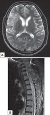
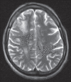
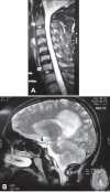
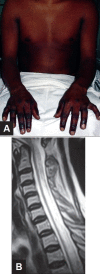
In hospitals in the tropics, the availability of magnetic resonance imaging (MRI) facilities in urban areas and especially in teaching institutions have resulted in white matter diseases being frequently reported in a variety of clinical settings. Unlike the west where multiple sclerosis (MS) is the commonest white matter disease encountered, in the tropics, there are myriad causes for the same. Infectious and post infectious disorders probably account for the vast majority of these diseases. Human immunodeficiency virus (HIV) infection tops the list of infective conditions. Central nervous system (CNS) tuberculosis occasionally presents with patchy parenchymal lesions unaccompanied by meningeal involvement. Human T cell leukemia virus (HTLV) infection and cystic inflammatory lesions such as neurocysticercosis are important causes to be considered in the differential diagnosis. Diagnosing post infectious demyelinating disorders is equally challenging since more than a third of cases seen in the tropics do not present with history of past infection or vaccinations. Metabolic and deficiency disorders such as Wernicke's encephalopathy, osmotic demyelinating syndrome associated with extra pontine lesions and Vitamin B12 deficiency states can occassionaly cause confusion in diagnosis. This review considers a few important disorders which manifest with white matter changes on MRI and create diagnostic difficulties in a population in the tropics.
Keywords: Demyelinating disorders; white matter diseases.
Conflict of interest statement
Figures


Similar articles
-
Seropositive Neuromyelitis Optica in a Case of Undiagnosed Ankylosing Spondylitis: A Neuro-Rheumatological Conundrum.Qatar Med J. 2022 Jul 7;2022(3):29. doi: 10.5339/qmj.2022.29. eCollection 2022. Qatar Med J. 2022. PMID: 35864917 Free PMC article.
-
Differential diagnosis of multiple sclerosis and other inflammatory CNS diseases.Mult Scler Relat Disord. 2020 Jan;37:101452. doi: 10.1016/j.msard.2019.101452. Epub 2019 Oct 15. Mult Scler Relat Disord. 2020. PMID: 31670010 Review.
-
White Matter Diseases with Radiologic-Pathologic Correlation.Radiographics. 2016 Sep-Oct;36(5):1426-47. doi: 10.1148/rg.2016160031. Radiographics. 2016. Update in: Radiographics. 2020 May-Jun;40(3):E4-E7. doi: 10.1148/rg.2020190204. PMID: 27618323 Updated. Review.
-
Common Clinical and Imaging Conditions Misdiagnosed as Multiple Sclerosis: A Current Approach to the Differential Diagnosis of Multiple Sclerosis.Neurol Clin. 2018 Feb;36(1):69-117. doi: 10.1016/j.ncl.2017.08.014. Neurol Clin. 2018. PMID: 29157405 Review.
-
[Chronic inflammatory demyelinating polyradiculoneuropathy associated with central nervous system involvement--as compared to multiple sclerosis].Rinsho Shinkeigaku. 1990 Sep;30(9):939-43. Rinsho Shinkeigaku. 1990. PMID: 2265502 Japanese.
Cited by
-
Novel Alanyl-tRNA Synthetase 2 Pathogenic Variants in Leukodystrophies.Front Neurol. 2019 Dec 17;10:1321. doi: 10.3389/fneur.2019.01321. eCollection 2019. Front Neurol. 2019. PMID: 31920941 Free PMC article.
-
Imaging and clinical properties of inflammatory demyelinating pseudotumor in the spinal cord.Neural Regen Res. 2013 Sep 15;8(26):2484-94. doi: 10.3969/j.issn.1673-5374.2013.26.010. Neural Regen Res. 2013. PMID: 25206559 Free PMC article.
-
Early-Onset Multiple Sclerosis With Frequent Relapses: A Challenging Diagnosis With a Less Favorable Prognosis.Cureus. 2021 Mar 18;13(3):e13963. doi: 10.7759/cureus.13963. Cureus. 2021. PMID: 33880297 Free PMC article.
-
Juvenile Alexander Disease: A Rare Leukodystrophy.Cureus. 2022 May 10;14(5):e24870. doi: 10.7759/cureus.24870. eCollection 2022 May. Cureus. 2022. PMID: 35698668 Free PMC article.
-
Rationalization of Using the MR Diffusion Imaging in B12 Deficiency.Ann Indian Acad Neurol. 2020 Jan-Feb;23(1):72-77. doi: 10.4103/aian.AIAN_485_18. Ann Indian Acad Neurol. 2020. PMID: 32055125 Free PMC article.
References
-
- Levy RM, Bredesen DE, Rosenbloom ML. Neurologic manifestations of the acquired immunodeficiency syndrome (AIDS): Experience at UCSF and review of the literature. J Neurosurg. 1985;62:475–95. - PubMed
-
- Petito CK, Cho ES, Lenman W, Navia BA, Price RW. Neuropathology of acquired immunodeficiency syndrome (AIDS): An autopsy review. J Neuropathol Exp Neurol. 1986;45:635–46. - PubMed
-
- Holland NR, Power C, Matthews VP, Glass VD, Forman M, McArthur JC. Cytomegalovirus encephalitis in acquired immunodeficiency syndrome (AIDS) Neurology. 1994;44:507–14. - PubMed
-
- Whiteman ML, Post JD, Berger JR, Tate LG, Bell MD, Limonte LP. Progressive multifocal leucoencephalopathy (PML) in 47 HIV-seropositive patients: Neuroimaging with clinical and pathologic correlation. Radiology. 1993;187:233–40. - PubMed
-
- Albrecht H, Hoffmann C, Degen O, Stoehr A, Plettenberg A, Mertenskötter T, et al. Highly active antiretroviral therapy significantly improves the prognosis of patients with HIV-associated progressive multifocal leucoencephalopathy. AIDS. 1998;12:1149–54. - PubMed
LinkOut - more resources
Full Text Sources
Miscellaneous

