. 2010 May;18(5):570-80.
doi: 10.1007/s00167-010-1064-x. Epub 2010 Feb 12.

Osteochondral defects in the ankle: why painful?

Affiliations

Osteochondral defects in the ankle: why painful?

C Niek van Dijk et al. Knee Surg Sports Traumatol Arthrosc. 2010 May.

Abstract

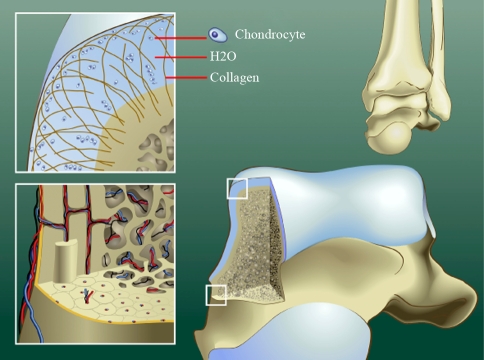
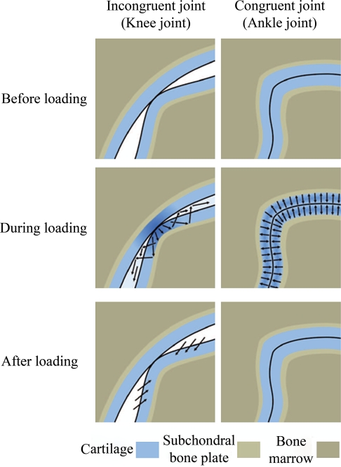
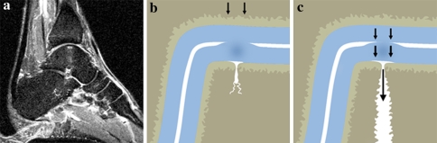
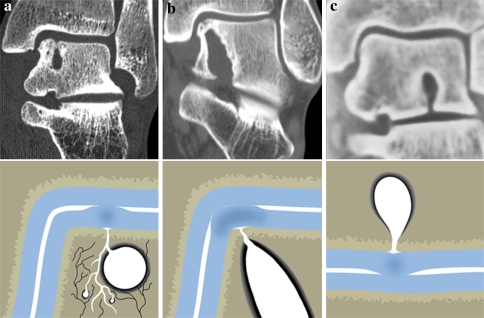
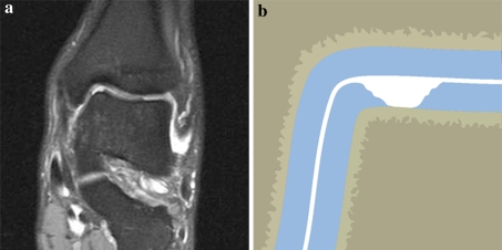
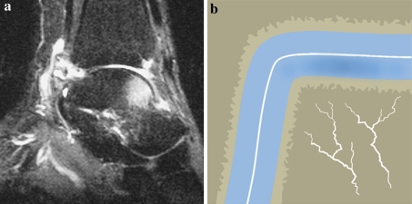
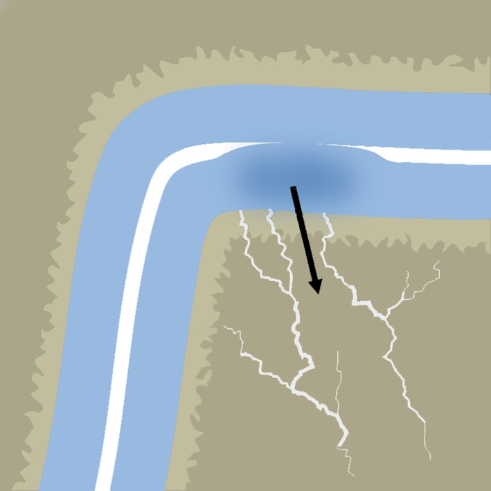
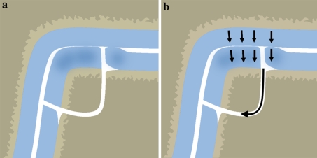
Osteochondral defects of the ankle can either heal and remain asymptomatic or progress to deep ankle pain on weight bearing and formation of subchondral bone cysts. The development of a symptomatic OD depends on various factors, including the damage and insufficient repair of the subchondral bone plate. The ankle joint has a high congruency. During loading, compressed cartilage forces its water into the microfractured subchondral bone, leading to a localized high increased flow and pressure of fluid in the subchondral bone. This will result in local osteolysis and can explain the slow development of a subchondral cyst. The pain does not arise from the cartilage lesion, but is most probably caused by repetitive high fluid pressure during walking, which results in stimulation of the highly innervated subchondral bone underneath the cartilage defect. Understanding the natural history of osteochondral defects could lead to the development of strategies for preventing progressive joint damage.


Figures

Fig. 1
Fig. 1
Schematic diagrams showing normal anatomy of ankle cartilage, subchondral plate and subchondral bone area. The cartilage consist of chondrocytes that lie groupwise in lacunae of the extracellular matrix, which contains collagen fibers in an arcwise configuration, hyaluronic acid, proteoglycans and 75% water (upper left). The hollow haversian canal that runs longitudinally down the center of the osteon in compact bone contains an arteriole, venule and lymphatic duct for vascular and lymphatic drainage. The Volkmann canals run perpendicular to and connect the Haversian canals (lower left)
Fig. 2
Fig. 2
Schematic diagrams showing the calculation of load transmission through the ankle joint during walking. Approximately one-sixth of the load across the ankle is transmitted through the talo-fibular facet, and the remaining load is transmitted through the tibiotalar articulation. F = force
Fig. 3
Fig. 3
Graph showing load in relation to tibiotalar contact (black line). The green line represents the average tibiotalar contact area of 4.4 cm² for a 75-kg person during the stance phase of walking. The blue line represents the same person with a tibiotalar contact area diminished by 42% to 2.6 cm², as would occur after an ankle fracture with 1 mm of lateral displacement of the talus and fibula. The red line represents the same person with a tibiotalar contact area diminished by 58% to 1.8 cm², as would occur after an ankle fracture with 2 mm of lateral displacement of the talus and fibula. The yellow line represents a person weighing 75 kg with an OD of the talus measuring 0.65 cm2; the average load on the remaining cartilage is increased from 650 to 764 N/cm²
Fig. 4
Fig. 4
Schematic comparison of the deformation of the cartilage in a congruent (ankle) and incongruent (knee) joint before, during and after loading. Arrows = direction of water
Fig. 5
Fig. 5
a Sagittal T2-weighted MRI study of an ankle with an OD. The vertical configuration of the water column (seen in the center of the talus) suggests that the water is pumped directly caudal under high pressure, perpendicular to the talar joint surface. b and c, Schematic diagrams of fissures in the subchondral bone plate of an unloaded ankle b and a loaded ankle c. When the ankle is loaded, the water is squeezed out of the cartilage into the subchondral bone. The diameter of the opening of the subchondral bone plate determines the pressure of the fluid flow (the smaller the diameter, the higher the pressure)
Fig. 6
Fig. 6
a through c, Coronal CT scans (upper row) with corresponding schematic diagrams (lower row), showing the ankles of three young patients (26–37 years), who had deep ankle pain of 5–12 years duration. An opening in the subchondral bone plate can be seen in all three CT scans, with subchondral osteolysis that has developed into a subchondral cyst. a Coronal CT, showing a cystic lesion in the talar body, with corresponding diagram schematically indicating the mechanism of cyst formation. Black lines = nerve endings in subchondral bone. b, In this patient, the cyst has extended to the subtalar joint. c, Sclerosis is visible around the subtalar cyst
Fig. 7
Fig. 7
a MRI study showing a cartilage defect of the medial talar dome. The subchondral bone plate has remained intact, and there is no sign of bone bruise. b Schematic diagram showing a fragment that probably was sheared from the underlying bone
Fig. 8
Fig. 8
a Sagittal T2-weighted MRI study of an ankle with a reticular bone bruise. The white area in the anterior talus represents bone edema. b Schematic diagram of a reticular bone bruise with intact subchondral bone plate. This type of bone bruise heals from the periphery to the center without complications
Fig. 9
Fig. 9
Schematic diagram showing the geographic type of bone bruise, which is continuous with the adjacent articular surface. Healing depends of the healing of the subchondral bone plate
Fig. 10
Fig. 10
Schematic diagrams showing a loose osteochondral fragment when the ankle is unloaded (a) and loaded (b). Healing under loading may be precluded by intermittent fluid flow around the fragment

Similar articles

Cited by

References

    1. Anderson DV, Lyne ED. Osteochondritis dissecans of the talus: case report on two family members. J Pediatr Orthop. 1984;4:356–357. - PubMed
    1. Aspenberg P, Van der Vis H. Migration, particles, and fluid pressure. A discussion of causes of prosthetic loosening. Clin Orthop Relat Res. 1998;352:75–80. doi: 10.1097/00003086-199807000-00010. - DOI - PubMed
    1. Astrand J, Skripitz R, Skoglund B, Aspenberg P. A rat model for testing pharmacologic treatments of pressure-related bone loss. Clin Orthop Relat Res. 2003;409:296–305. doi: 10.1097/01.blo.0000052938.71325.46. - DOI - PubMed
    1. Berndt AL, Harty M. Transchondral fractures (osteochondritis dissecans) of the talus. J Bone Joint Surg Am. 1959;41-A:988–1020. - PubMed
    1. Braune W, Fischer O. Die Bewegungen des Kniegelenks nach einer neuen Methode am lebenden Menschen gemessen. Leipzig: S Hirzel; 1891. pp. 75–150.

MeSH terms