. 2010 Feb 12:10:15.
doi: 10.1186/1471-213X-10-15.

BMP-2 functions independently of SHH signaling and triggers cell condensation and apoptosis in regenerating axolotl limbs

Affiliations

BMP-2 functions independently of SHH signaling and triggers cell condensation and apoptosis in regenerating axolotl limbs

Jean-Charles Guimond et al. BMC Dev Biol. .

Abstract

Background: Axolotls have the unique ability, among vertebrates, to perfectly regenerate complex body parts, such as limbs, after amputation. In addition, axolotls pattern developing and regenerating autopods from the anterior to posterior axis instead of posterior to anterior like all tetrapods studied to date. Sonic hedgehog is important in establishing this anterior-posterior axis of limbs in all tetrapods including axolotls. Interestingly, its expression is conserved (to the posterior side of limb buds and blastemas) in axolotl limbs as in other tetrapods. It has been suggested that BMP-2 may be the secondary mediator of sonic hedgehog, although there is mounting evidence to the contrary in mice. Since BMP-2 expression is on the anterior portion of developing and regenerating limbs prior to digit patterning, opposite to the expression of sonic hedgehog, we examined whether BMP-2 expression was dependent on sonic hedgehog signaling and whether it affects patterning of the autopod during regeneration.

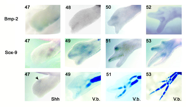
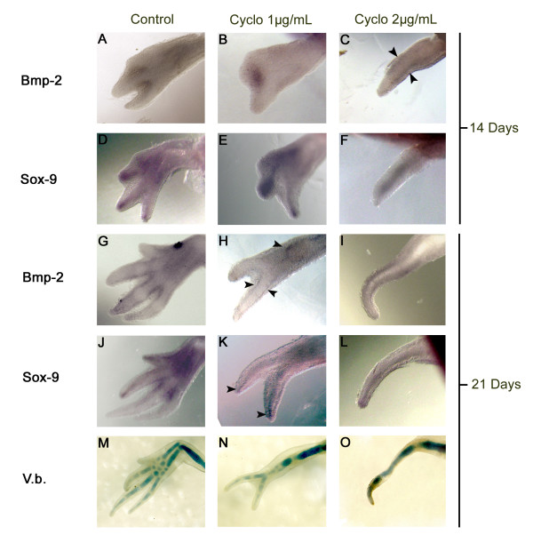
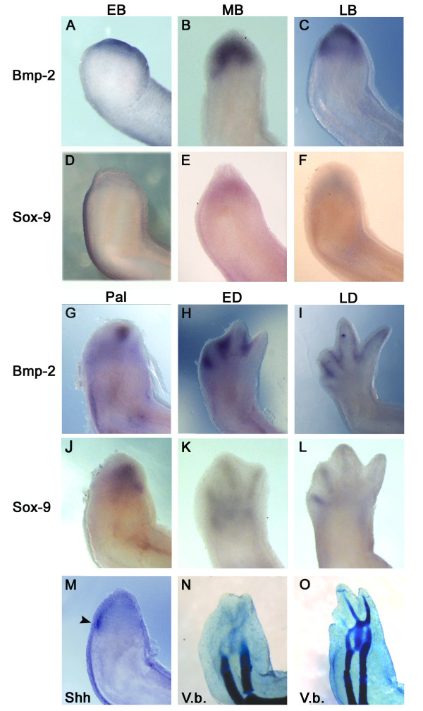
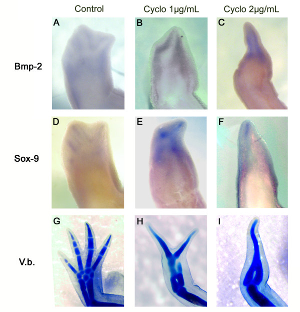
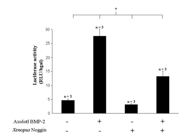
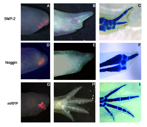
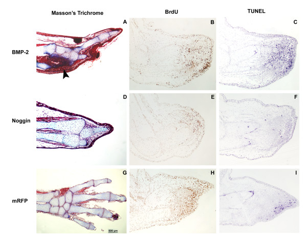
Results: The expression of BMP-2 and SOX-9 in developing and regenerating axolotl limbs corresponded to the first digits forming in the anterior portion of the autopods. The inhibition of sonic hedgehog signaling with cyclopamine caused hypomorphic limbs (during development and regeneration) but did not affect the expression of BMP-2 and SOX-9. Overexpression of BMP-2 in regenerating limbs caused a loss of digits. Overexpression of Noggin (BMP inhibitor) in regenerating limbs also resulted in a loss of digits. Histological analysis indicated that the loss due to BMP-2 overexpression was the result of increased cell condensation and apoptosis while the loss caused by Noggin was due to a decrease in cell division.

Conclusion: The expression of BMP-2 and its target SOX-9 was independent of sonic hedgehog signaling in developing and regenerating limbs. Their expression correlated with chondrogenesis and the appearance of skeletal elements has described in other tetrapods. Overexpression of BMP-2 did not cause the formation of extra digits, which is consistent with the hypothesis that it is not the secondary signal of sonic hedgehog. However, it did cause the formation of hypomorphic limbs as a result of increased cellular condensation and apoptosis. Taken together, these results suggest that BMP-2 does not have a direct role in patterning regenerating limbs but may be important to trigger condensation prior to ossification and to mediate apoptosis.


Figures

Figure 1
Figure 1
Expression patterns (whole mount in situ hybridisation) for BMP-2, SOX-9 and Shh in developing limbs and cartilage staining (Victoria blue V.b.). All pictures show a dorsal view of the limb with the posterior side on top. Numbers in the upper left of each panel refers to the developmental stages [51].
Figure 2
Figure 2
Expression of BMP-2 (A-C) and SOX-9 (D-F) detected by whole mount in situ hybridisation in developing limbs treated 14 days with cyclopamine. Expression of BMP-2 (G-I) and SOX-9 (J-L) in developing limbs treated 21 days with cyclopamine. Left column (A, D, G, J, M) shows control animals (0.4 μL EtOH/mL), middle column (B, E, H, K, N) shows the animals treated with 1 μg/mL of cyclopamine and right column (C, F, I, L, O) shows the animals treated with 2 μg/mL of cyclopamine. All pictures show a dorsal view of the limbs with posterior side on top. Arrowheads in C, H and K indicate the expression of BMP-2 on both sides of the regenerating digits and the expression of SOX-9 in the middle of the regenerating digits. The last row (M-O) shows the cartilage staining (Victoria blue) of the skeletal elements at the end of cyclopamine treatments (21 days).
Figure 3
Figure 3
Expression pattern, by whole mount in situ hybridisation, for BMP-2 (A-C and G-I), SOX-9 (D-F and J-L), Shh (M) and cartilage staining of skeletal elements (Victoria blue; N-O) at different stages of limb regeneration. All pictures show a dorsal view of the regenerating limbs with posterior side on the left. EB, early bud; MB, medium bud; LB, late bud; Pal, palette; ED, early differentiation; LD, late differentiation. The arrowhead in panel M indicates the restricted expression of Shh in the posterior portion of the blastema.
Figure 4
Figure 4
Expression pattern, by whole mount in situ hybridisation, for BMP-2 (A-C) and SOX-9 (D-F) at the ED stage in regenerating limb treated with cyclopamine from the time of amputation until fixed at ED. Left column (A, D, G) shows control animals (treated with the carrier 0.4 μL EtOH/mL), middle column (B, E, H) shows the animals treated with 1 μg/mL of cyclopamine and right column (C, F, I) shows the animals treated with 2 μg/mL of cyclopamine. All pictures show a dorsal view of the limbs with the posterior side on the left. The bottom row (G, H, I) show the skeletal staining (Victoria blue) at the end of the treatments when the control animals had completely regenerated.
Figure 5
Figure 5
Activation of BMP responsive promoter construct by ectopic expression of axolotl BMP-2. The graphic summarizes BMP responsive luciferase activity performed in the human chondrocyte cell line, C28/I2, after the transfection of BMP-2, Noggin or BMP-2 and Noggin expression plasmids combined. For each condition, the BMP responsive reporter construct and the β-galactosidase reporter construct were always co-transfected. The first bar shows the basal luciferase activity (transfected with mRFP without axolotl BMP-2 or Xenopus laevis Noggin) of 4.6 ± 0.6. The second bar shows a significant increase (27.7 ± 3.4) in luciferase activity when the axolotl BMP-2 expressing plasmid was transfected. The third bar indicates that transfection of the Xenopus laevis Noggin expressing plasmid decreased luciferase activity (3.1 ± 0.3) compared to basal activity. The fourth bar shows the luciferase activity from the co-transfection of the axolotl BMP-2 and the Xenopus laevis Noggin expressing plasmids. This co-expression resulted in a significant decrease in luciferase activity (13.3 ± 1.9) compared to the expression of BMP-2 alone. All results (mean ± sd; n = 3/condition) are expressed as relative light unit (RLU) normalised with β-galactosidase activity.
Figure 6
Figure 6
Ectopic expression of axolotl BMP-2 and Xenopus laevis Noggin by electroporating the expression plasmids directly into regenerating limbs. Different plasmid combinations were transfected to test the effect of BMP-2 and Noggin on the regeneration process. In order to assess the efficiency of the electroporation in vivo (first column A, D, G), the mRFP expressing plasmid was co-transfected with axolotl BMP-2 (A), Xenopus laevis Noggin (D) or mRFP alone (G; control) in the posterior half of regenerating blastema at the LB stage. The second column (B, E, H) show the resulting phenotypes once the controls (mRFP ectopic expression alone) were fully regenerated and the third column (C, F, I) show their skeletal elements using Victoria blue staining.
Figure 7
Figure 7
Histological analysis, cellular proliferation and apoptosis of regenerating limbs transfected with axolotl BMP-2, Xenopus laevis Noggin and mRFP. Ectopic expression of axolotl BMP-2 (A-C), Xenopus laevis Noggin (D-F) and mRFP (G-I) by electroporating the expression plasmids directly into regenerating limbs were further analysed at the cellular level. Panels A, D and G show the resulting phenotypes at the tissue level on 10 μm sections with the Masson's trichrome staining. Clear differences can be observed between the ectopic expression of axolotl BMP-2 (A: condensation area pointed out by the black arrow), Xenopus laevis Noggin (D: no condensation visible) and the mRFP control (G, normal regenerate). Panels B, E and H show the level of BrdU incorporation as a measure of cell division. Overexpression of BMP-2 6 days after electroporation (B) did not seem to affect cell division compared to mRFP control (H). However, Noggin overexpression significantly reduced the number of BrdU positive cells 6 days after electroporation, especially on the posterior side where it was overexpressed (E). Panels C, F and I show the level of apoptosis as determined by TUNEL assay. BMP-2 overexpression (C) significantly increased the number of apoptotic cells during regeneration compared to Noggin (F), which reduced apoptosis, and mRFP control (I).

Similar articles

Cited by

References

    1. Urist MR. Bone: formation by autoinduction. Science. 1965;150:893–9. doi: 10.1126/science.150.3698.893. - DOI - PubMed
    1. Wozney JM. Overview of bone morphogenetic proteins. Spine. 2002;27:S2–8. doi: 10.1097/00007632-200208151-00002. - DOI - PubMed
    1. Munoz-Sanjuan I, Brivanlou AH. Neural induction, the default model and embryonic stem cells. Nat Rev Neurosci. 2002;3:271–80. doi: 10.1038/nrn786. - DOI - PubMed
    1. Suzuki A, Kaneko E, Ueno N, Hemmati-Brivanlou A. Regulation of epidermal induction by BMP2 and BMP7 signaling. Dev Biol. 1997;189:112–22. doi: 10.1006/dbio.1997.8652. - DOI - PubMed
    1. Riddle RD, Johnson RL, Laufer E, Tabin C. Sonic hedgehog mediates the polarizing activity of the ZPA. Cell. 1993;75:1401–16. doi: 10.1016/0092-8674(93)90626-2. - DOI - PubMed

Publication types