. 2010 Feb 12:9:37.
doi: 10.1186/1476-4598-9-37.

Sindbis viral vector induced apoptosis requires translational inhibition and signaling through Mcl-1 and Bak

Affiliations

Sindbis viral vector induced apoptosis requires translational inhibition and signaling through Mcl-1 and Bak

Lisa Venticinque et al. Mol Cancer. .

Abstract

Background: Sindbis viral vectors are able to efficiently target and kill tumor cells in vivo, as shown using pancreatic and ovarian cancer models. Infection results in apoptosis both in vitro and in vivo. Sindbis vector uptake is mediated by the LAMR, which is upregulated on a number of different tumor types, thus conferring specificity of the vector to a wide range of cancers. In this study we elucidate the mechanism of apoptosis in two tumor cell lines, MOSEC, derived from the ovarian epithelium and Pan02, derived from a pancreatic adenocarcinoma. A comprehensive understanding of the mechanism of apoptosis would facilitate the design of more effective vectors for cancer therapy.

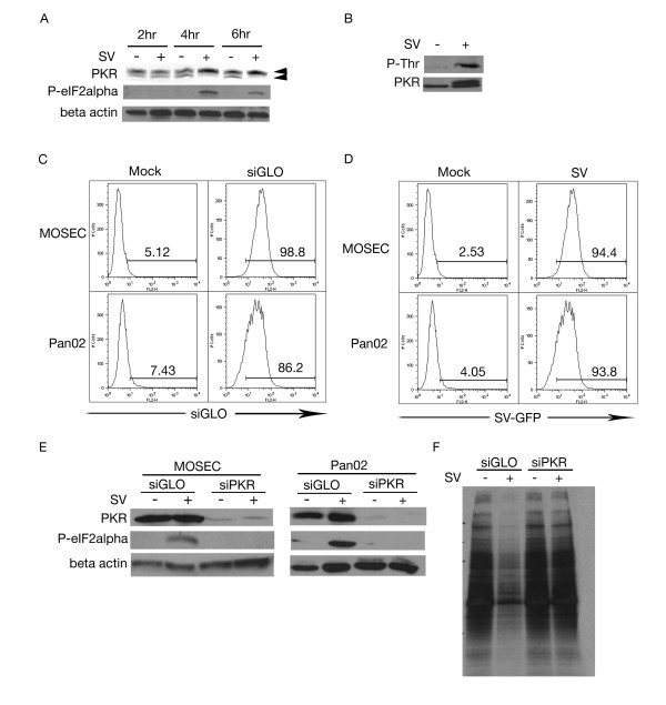
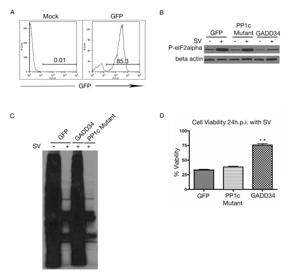
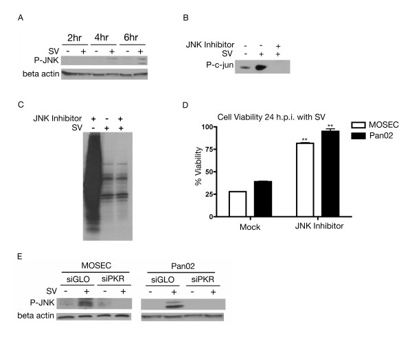
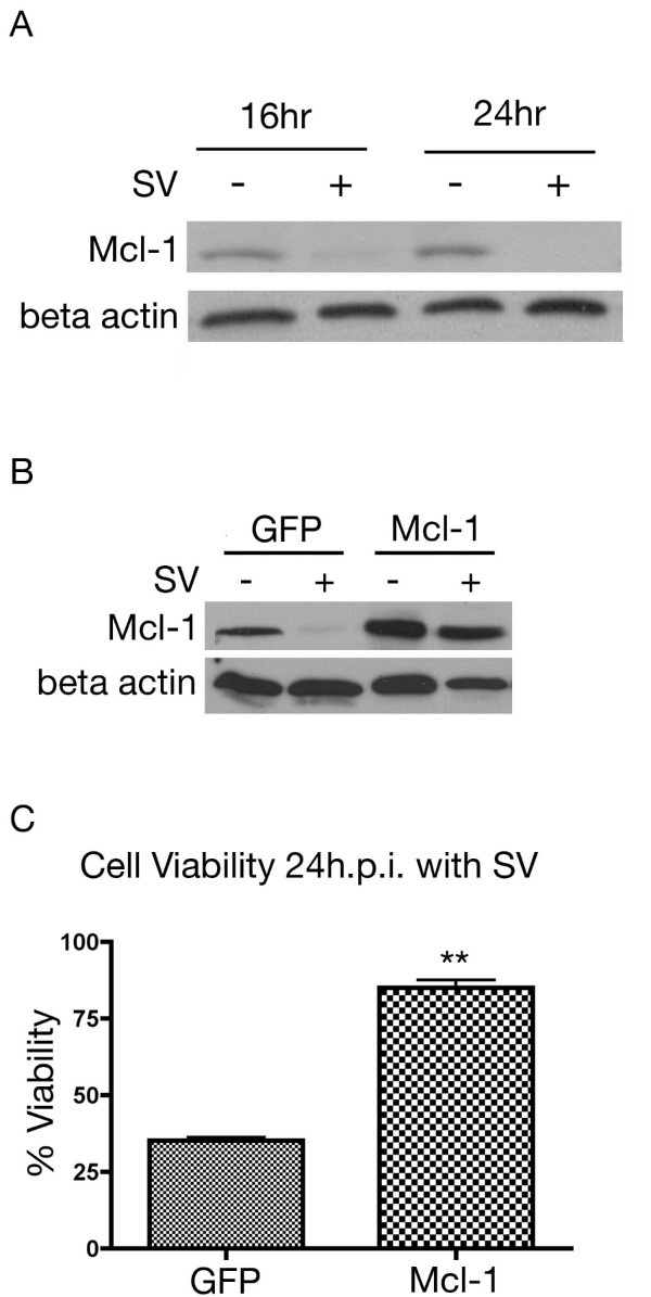
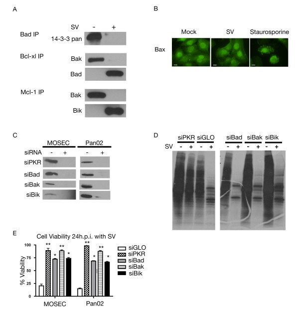
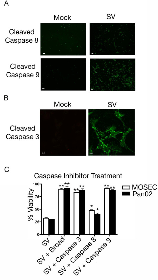
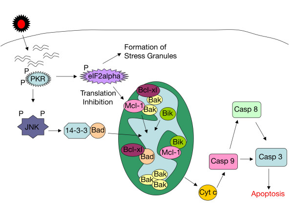
Results: The initial phase of Sindbis vector induced apoptosis in MOSEC and Pan02 models reconfirms that viral infection is sensed by PKR due to double-stranded RNA intermediates associated with genomic replication. PKR activation results in translation inhibition through eIF2alpha phosphorylation and initiation of the stress response. Our studies indicate that the roles of two proteins, Mcl-1 and JNK, intimately link Sindbis induced translational arrest and cellular stress. Translational arrest inhibits the synthesis of anti-apoptotic Bcl-2 protein, Mcl-1. JNK activation triggers the release of Bad from 14-3-3, which ultimately results in apoptosis. These signals from translational arrest and cellular stress are propagated to the mitochondria where Bad and Bik bind to Bcl-xl and Mcl-1 respectively. Formation of these heterodimers displaces Bak, which results in caspase 9 cleavage and signaling through the mitochondrial pathway of apoptosis.

Conclusion: The host cell response to Sindbis is triggered through PKR activation. Our studies demonstrate that PKR activation and subsequent translational arrest is linked to both cellular stress and apoptosis. We have also found the linkage point between translational arrest and apoptosis to be Mcl-1, a protein whose constant translation is required for inhibition of apoptosis. With this information vectors can be designed, which express or repress proteins implicated in this study, to enhance their therapeutic potential.


Figures

Figure 1
Figure 1
dsRNA species activate PKR. (A) Sindbis vector (SV) infection leads to activation of PKR. Lysates collected at 2, 4 and 6 h.p.i. from mock (-) or SV infected (+) MOSEC cells were analyzed by immunoblotting using a polyclonal antibody for PKR (upper arrow represents the phosphorylated form, lower arrow, non-phosphorylated form). (B) PKR is threonine phosphorylated following infection. Lysates were subjected to immunoprecipitation with PKR and blotted for phospho-threonine. SV infected MOSEC cells but not mock infected cells show phosphorylation indicating that this event is the result of infection. (C) MOSEC and Pan02 cells are efficiently transfected with siRNA. MOSEC and Pan02 cells were transfected with siGLO, a fluorescently conjugated control siRNA. Cells were collected and subjected to FACS analysis. (D) SV-GFP efficiently infects both MOSEC and Pan02 cells. 16 h.p.i. with SV-GFP, cells were harvested for FACS analysis monitoring GFP expression. (E) eIF2α phosphorylation is inhibited in cells with diminished PKR expression. Lysates were collected from MOSEC or Pan02 cells, transfected with siPKR or siGLO and infected with SV or mock infected, at 6 h.p.i. Immunoblotting for phospho-eIF2α indicates a lack of phosphorylation in siPKR-transfected cells. Immunoblots in panels A and E were stripped and reprobed with β actin for loading control. (F) Transfection of siPKR inhibits Sindbis-induced translational arrest. MOSEC cells transfected with either siPKR or siGLO were infected with SV or mock infected and subjected to 35S labeling. Decreased translation was observed in the siGLO transfected/SV infected sample however not the similarly treated siPKR sample.
Figure 2
Figure 2
Translational arrest is inhibited by ablation of PKR expression. (A) MOSEC cells are efficiently transfected with GFP control vector. At 48 hours post transfection MOSEC cell were harvested for FACS analysis monitoring FL-1 for GFP expression. (B) Cells transfected with GADD34, but not the GADD34 PP1c deletion mutant, can dephosphorylate eIF2α. MOSEC cells transfected with GADD34, PP1c mutant or GFP control vector were infected at 48 hours. 8 h.p.i. lysates were collected and subjected to western blotting for phospho-eIF2α. In panel B immunoblot was stripped and reprobed with β actin for loading control. (C) Transfection of GADD34 but not the PP1c mutant alleviates Sindbis vector-induced translational arrest. MOSEC cells transfected with GADD34, PP1c mutant or GFP control vector were infected at 48 hours. 24 h.p.i. cells were subjected to 35S labeling. (D) Dephosphorylation of eIF2α by GADD34 but not the GADD34 mutant lacking the PP1c interacting domain, increases the cell viability at 24 h.p.i. MOSEC cells were transfected with either GADD34, GADD34 PP1c mutant or GFP control vector. 48 hours after transfection cells were infected with SV. 24 h.p.i. cell viability was assessed. Data in D represents the SEM (error bars) of three experiments. Cell viability for each sample was compared to the infected control at the same time point and was corrected for the percentage of infected cells. Statistical significance was calculated by a two-tailed student t-test (** P < 0.005).
Figure 3
Figure 3
Sindbis vector infection induces the formation of stress granules. (A) Infection with Sindbis vector induces the formation of stress granules, which can be inhibited by siPKR transfection. MOSEC cells transfected with siPKR or siGLO and infected with SV or mock infected were processed for immunofluorescence for stress granule marker TIA-1 at 6 h.p.i. Arrows in each panel indicate stress granules. (B&C) Sindbis vector infection results in the sequestration of translation initiation factors in stress granules. MOSEC cells were infected with SV or mock infected and processed for immunofluorescence using antibodies for TIA-1 and eIF4E (B) or eIF4G (C) at 6 h.p.i. Co-localization of translation initiation factors with TIA-1 indicates that they are located in stress granules following infection. Scale bars for panels A-C indicate 10 μm.
Figure 4
Figure 4
Sindbis vector infection activates JNK. (A) JNK phosphorylation is induced by infection with Sindbis vector. Lysates of SV (+) infected or mock (-) infected MOSEC cells were collected at 2, 4 and 6 h.p.i. Immunoblotting with phospho-JNK first detects phosphorylation at 4 h.p.i. (B) Cell permeable JNK inhibitory peptide inhibits c-jun phosphorylation. MOSEC cells were treated for 1 hour with JNK specific inhibitory peptide and then infected with SV or mock infected and subjected to a JNK specific kinase assay 24 h.p.i. (C) Inhibition of JNK activation has no effect on Sindbis-induced translational arrest. MOSEC cells treated with JNK inhibitor and infected were labeled with 35S methionine 24 h.p.i. (D) JNK inhibition results in an increase in cell viability following Sindbis infection. MOSEC or Pan02 cells pretreated for 1 hour with a JNK-specific peptide inhibitor and then infected with SV or mock infected were assessed for cell viability 24 h.p.i. Data in D represents the SEM (error bars) of three experiments. Each sample was compared to the non-treated infected control at the same time point. Statistical significance was calculated by a two-tailed student t-test (** P < 0.005). (E) JNK phosphorylation is downstream of PKR activation. Lysates from MOSEC or Pan02 cells, transfected with siPKR or siGLO (control) and infected with SV or mock infected, were collected at 8 h.p.i. Immunoblotting for phosphorylated JNK indicates that it remains dephosphorylated in cells treated with siPKR. The immunoblots in A&E were stripped and reprobed for β actin as a loading control.
Figure 5
Figure 5
Role of Mcl-1 in Sindbis infection. (A) Sindbis vector infection induces the degradation of Mcl-1. Lysates were collected from MOSEC cells infected with SV (+) or mock (-) infected. Immunoblot analysis indicates a loss of Mcl-1 expression by 16 h.p.i. (B) MOSEC cells transfected with Mcl-1 vector overexpress the protein. Cells transfected with Mcl-1 or GFP control vector were infected at 48 hours post transfection. At 24 h.p.i. lysates were collected and subjected to western blotting for Mcl-1. In A and B immunoblots were stripped and reprobed with β actin for loading control. (C) Mcl-1 overexpression protects the cells from Sindbis-induced loss of viability. MOSEC cells were transfected with Mcl-1 or GFP transfected. At 48 hours cells were infected with Sindbis vector. 24 h.p.i. cell viability was assessed. Data in C represents the SEM (error bars) of three experiments. Each sample was compared to the infected control at the same time point. Statistical significance was calculated by a two-tailed student t-test (** P < 0.005).
Figure 6
Figure 6
Involvement of Bcl-2 proteins in the cellular response. (A) Infection with Sindbis results in a shift in anti-apoptotic Bcl-2 family heterodimer composition. Cell lysates were collected from SV (+) or mock (-) infected MOSEC cells 20 h.p.i. and subjected to mitochondrial isolation. Fractionated lysates were immunoprecipitated with antibodies specific to Bad (cytoplasmic fraction), Bcl-xl and Mcl-1 (mitochondrial fraction). (B) Bax remains in the cytoplasm following infection with Sindbis vector. MOSEC cells were infected with SV-Luc or treated with staurosporine, as positive control. At 20 h.p.i. samples were subjected to immunofluorescence staining for Bax. Scale bars indicate 10 μm. (C) Knockdown of proteins using targeted siRNA is significant. MOSEC or Pan02 cells were transfected with indicated siRNAs. Lysates were collected 24 h.p.i. and subjected to western blotting for indicated proteins. (D) Translation is still inhibited in cells after protein knockdown. MOSEC cells transfected with siRNA directed against indicated proteins and infected with SV or mock infected were subjected to 35S methionine labeling 24 h.p.i. (E) Ablation of expression of certain BH3 only proteins can partially protect cells from Sindbis vector infection. MOSEC or Pan02 cells were treated with indicated siRNAs (or siGLO control) and infected with Sindbis vector. Cell viability was assessed 24 h.p.i. Data in E represents the SEM (error bars) of three experiments. Cell viability for each sample was compared to the infected control and corrected for percentage of infection. Statistical significance was calculated by a two-tailed student t-test (* P < 0.05, ** P < 0.005).
Figure 7
Figure 7
Sindbis vector infection causes caspase cleavage. (A) Sindbis vector infection leads to significant cleavage of caspase 9 but minimal amounts of caspase 8. MOSEC cells infected with SV or mock infected were assayed 24 h.p.i. for caspase activation using fluorescent probes specific for individual caspase cleavage products. Scale bars for panel A indicate 100 μm. (B) Infection with Sindbis viral vector results in the cleavage of caspase 3. MOSEC cells infected with SV or mock infected were assayed at 24 h.p.i. for caspase 3 cleavage using a cleavage specific fluorescent probe. Scale bars indicate 10 μm. (C) Treatment with selective caspase inhibitors can protect cells from a loss of cell viability. MOSEC or Pan02 cells were infected with Sindbis vector or mock infected and then incubated with caspase specific peptide inhibitors. Cell viability was assessed at 24 h.p.i. Cell viability indicates that cells treated with a broad caspase inhibitor, caspase 3 inhibitor or caspase 9 inhibitor are protected from apoptosis whereas samples treated with caspase 8 inhibitor were not. Data in C represents the SEM (error bars) of three experiments. Each sample was compared to the infected control at the same time point. Statistical significance was calculated by a two-tailed student t-test (* P < 0.05, ** P < 0.005).
Figure 8
Figure 8
Schematic diagram of the cellular response to Sindbis vector infection. Sindbis vector replication forms double-stranded RNA species, which activate PKR. PKR is able to inhibit cellular translation through eIF2α phosphorylation and the formation of stress granules. Translation inhibition blocks the production of new Mcl-1, which is rapidly turned over. The remaining Mcl-1, in complex with Bak, is displaced by BH3 only protein Bik, thus releasing Bak. Downstream of PKR, JNK is activated, which causes the release of Bad from its complex with 14-3-3. Bad translocates to the mitochondria and disrupts the complex between Bcl-xl and Bak. Bak oligomerizes resulting in the release of cytochrome c from the mitochondria. Caspase 9 is cleaved causing caspase 3 cleavage and downstream apoptosis.

Similar articles

Cited by

References

    1. Bast RC Jr, Hennessy B, Mills GB. The biology of ovarian cancer: new opportunities for translation. Nat Rev Cancer. 2009;9(6):415–428. doi: 10.1038/nrc2644. - DOI - PMC - PubMed
    1. Freitas D, Fernandes Gdos S, Hoff PM, Cunha JE. Medical management of pancreatic adenocarcinoma. Pancreatology. 2009;9(3):223–232. doi: 10.1159/000199433. - DOI - PubMed
    1. Strauss JHaS EG. The Alphaviruses: Gene Expression, Replication, and Evolution. Microbiological Reviews. 1994;58(3):491–562. - PMC - PubMed
    1. Tseng JC, Hurtado A, Yee H, Levin B, Boivin C, Benet M, Blank SV, Pellicer A, Meruelo D. Using sindbis viral vectors for specific detection and suppression of advanced ovarian cancer in animal models. cancer research. 2004;64:6684–6692. doi: 10.1158/0008-5472.CAN-04-1924. - DOI - PubMed
    1. Tseng JC, Levin B, Hurtado A, Yee H, Perez de Castro I, Jiminez M, Shamamian P, Jin R, Novick RP, Pellicer A, Meruelo D. Systemic tumor targeting and killing by sindbis viral vectors. Nature Biotechnology. 2004;22(1):70–77. doi: 10.1038/nbt917. - DOI - PubMed

Publication types

MeSH terms

Substances