

Quantitative evaluation of motor function before and after engraftment of dopaminergic neurons in a rat model of Parkinson's disease
- PMID: 20152049
- PMCID: PMC2838763
- DOI: 10.1186/1423-0127-17-9
Quantitative evaluation of motor function before and after engraftment of dopaminergic neurons in a rat model of Parkinson's disease
Abstract
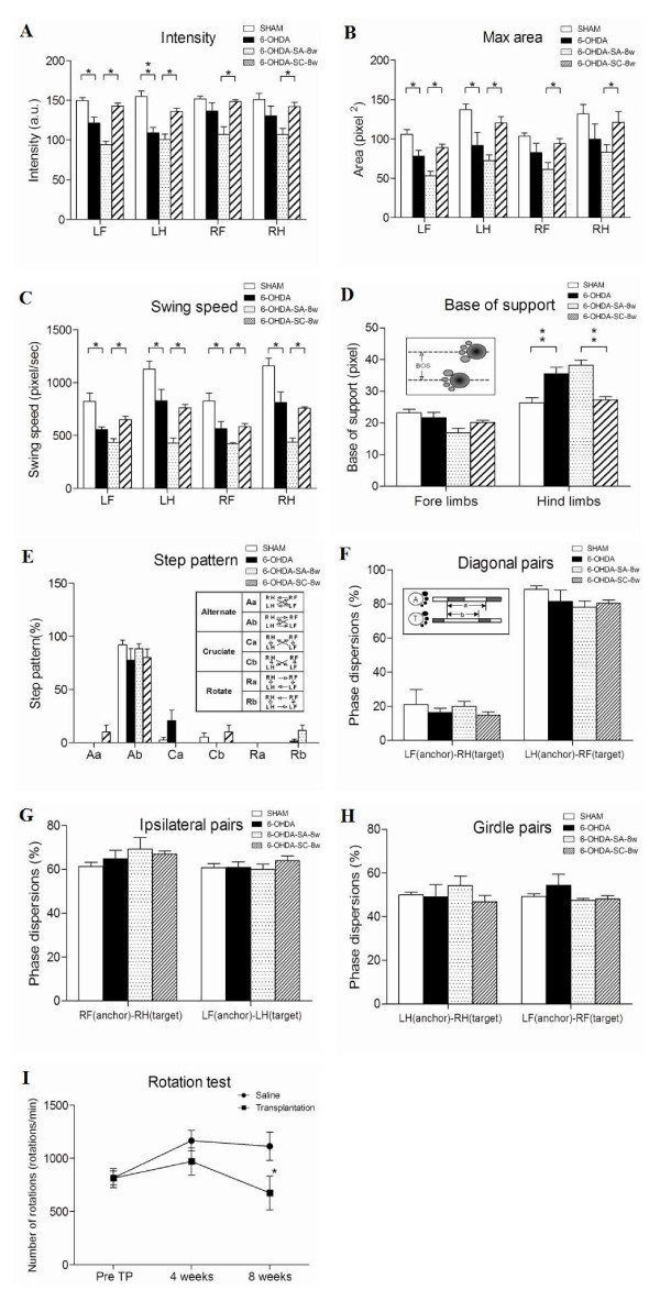
Although gait change is considered a useful indicator of severity in animal models of Parkinson's disease, systematic and extensive gait analysis in animal models of neurological deficits is not well established. The CatWalk-assisted automated gait analysis system provides a comprehensive way to assess a number of dynamic and static gait parameters simultaneously. In this study, we used the Catwalk system to investigate changes in gait parameters in adult rats with unilateral 6-OHDA-induced lesions and the rescue effect of dopaminergic neuron transplantation on gait function. Four weeks after 6-OHDA injection, the intensity and maximal area of contact were significantly decreased in the affected paws and the swing speed significantly decreased in all four paws. The relative distance between the hind paws also increased, suggesting that animals with unilateral 6-OHDA-induced lesions required all four paws to compensate for loss of balance function. At 8 weeks post-transplantation, engrafted dopaminergic neurons expressed tyrosine hydroxylase. In addition, the intensity, contact area, and swing speed of the four limbs increased and the distance between the hind paws decreased. Partial recovery of methamphetamine-induced rotational response was also noted.
Figures


Similar articles
-
Dopaminergic neuroprotection and regeneration by neurturin assessed by using behavioral, biochemical and histochemical measurements in a model of progressive Parkinson's disease.Brain Res. 2002 Aug 30;947(2):271-83. doi: 10.1016/s0006-8993(02)02934-7. Brain Res. 2002. PMID: 12176170
-
High frequency stimulation of the subthalamic nucleus improves speed of locomotion but impairs forelimb movement in Parkinsonian rats.Neuroscience. 2007 Sep 7;148(3):815-23. doi: 10.1016/j.neuroscience.2007.06.043. Epub 2007 Jul 17. Neuroscience. 2007. PMID: 17706885
-
Delayed gene therapy of glial cell line-derived neurotrophic factor is efficacious in a rat model of Parkinson's disease.Brain Res Mol Brain Res. 2005 Mar 24;134(1):155-61. doi: 10.1016/j.molbrainres.2004.06.029. Brain Res Mol Brain Res. 2005. PMID: 15790539
-
Walking pattern analysis after unilateral 6-OHDA lesion and transplantation of foetal dopaminergic progenitor cells in rats.Behav Brain Res. 2009 May 16;199(2):317-25. doi: 10.1016/j.bbr.2008.12.007. Epub 2008 Dec 13. Behav Brain Res. 2009. PMID: 19124044
-
Neuroprotective effects of erythropoietin on 6-hydroxydopamine-treated ventral mesencephalic dopamine-rich cultures.Exp Cell Res. 2010 Mar 10;316(5):737-46. doi: 10.1016/j.yexcr.2010.01.001. Epub 2010 Jan 11. Exp Cell Res. 2010. PMID: 20060824
Cited by
-
Targeted delivery of neural stem cells to the brain using MRI-guided focused ultrasound to disrupt the blood-brain barrier.PLoS One. 2011;6(11):e27877. doi: 10.1371/journal.pone.0027877. Epub 2011 Nov 16. PLoS One. 2011. PMID: 22114718 Free PMC article.
-
Step Sequence Is a Critical Gait Parameter of Unilateral 6-OHDA Parkinson's Rat Models.Cell Transplant. 2017 Apr 13;26(4):659-667. doi: 10.3727/096368916X693059. Epub 2016 Sep 26. Cell Transplant. 2017. PMID: 27677974 Free PMC article.
-
Cerebellar transcriptional alterations with Purkinje cell dysfunction and loss in mice lacking PGC-1α.Front Cell Neurosci. 2015 Jan 6;8:441. doi: 10.3389/fncel.2014.00441. eCollection 2014. Front Cell Neurosci. 2015. PMID: 25610371 Free PMC article.
-
Characterization of graded 6-Hydroxydopamine unilateral lesion in medial forebrain bundle of mice.Sci Rep. 2024 Feb 14;14(1):3721. doi: 10.1038/s41598-024-54066-0. Sci Rep. 2024. PMID: 38355892 Free PMC article.
-
Carnosic acid protects non-alcoholic fatty liver-induced dopaminergic neuron injury in rats.Metab Brain Dis. 2017 Apr;32(2):483-491. doi: 10.1007/s11011-016-9941-8. Epub 2016 Dec 13. Metab Brain Dis. 2017. PMID: 27957651
References
-
- Sidaway B, Anderson J, Danielson G, Martin L, Smith G. Effects of long-term gait training using visual cues in an individual with Parkinson disease. Phys Ther. 2006;86:186–194. - PubMed
Publication types
MeSH terms
Substances
LinkOut - more resources
Full Text Sources
Medical
