

Reverse micelle encapsulation of membrane-anchored proteins for solution NMR studies
- PMID: 20152148
- PMCID: PMC2876244
- DOI: 10.1016/j.str.2009.11.010
Reverse micelle encapsulation of membrane-anchored proteins for solution NMR studies
Abstract
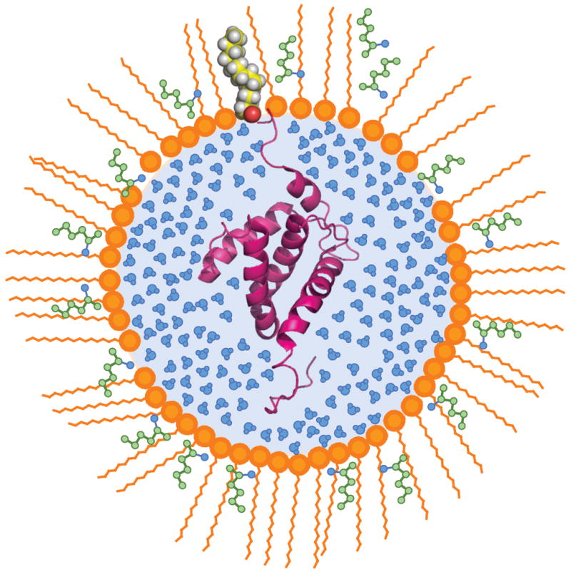
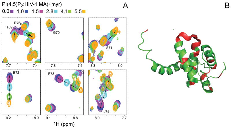
Perhaps 5%-10% of proteins bind to membranes via a covalently attached lipid. Posttranslational attachment of fatty acids such as myristate occurs on a variety of viral and cellular proteins. High-resolution information about the nature of lipidated proteins is remarkably sparse, often because of solubility problems caused by the exposed fatty acids. Reverse micelle encapsulation is used here to study two myristoylated proteins in their lipid-extruded states: myristoylated recoverin, which is a switch in the Ca(2+) signaling pathway in vision, and the myristoylated HIV-1 matrix protein, which is postulated to be targeted to the plasma membrane through its binding to phosphatidylinositol-4,5-bisphosphate. Both proteins have been successfully encapsulated in the lipid-extruded state and high-resolution NMR spectra obtained. Both proteins bind their activating ligands in the reverse micelle. This approach seems broadly applicable to membrane proteins with exposed fatty acid chains that have eluded structural characterization by conventional approaches.
Figures
Similar articles
-
Myristate exposure in the human immunodeficiency virus type 1 matrix protein is modulated by pH.Biochemistry. 2010 Nov 9;49(44):9551-62. doi: 10.1021/bi101245j. Biochemistry. 2010. PMID: 20886905 Free PMC article.
-
A method for solution NMR structural studies of large integral membrane proteins: reverse micelle encapsulation.Biochim Biophys Acta. 2010 Feb;1798(2):150-60. doi: 10.1016/j.bbamem.2009.07.027. Epub 2009 Aug 8. Biochim Biophys Acta. 2010. PMID: 19665988 Free PMC article. Review.
-
Trio engagement via plasma membrane phospholipids and the myristoyl moiety governs HIV-1 matrix binding to bilayers.Proc Natl Acad Sci U S A. 2013 Feb 26;110(9):3525-30. doi: 10.1073/pnas.1216655110. Epub 2013 Feb 11. Proc Natl Acad Sci U S A. 2013. PMID: 23401539 Free PMC article.
-
High-resolution NMR spectroscopy of encapsulated proteins dissolved in low-viscosity fluids.J Magn Reson. 2014 Apr;241:137-47. doi: 10.1016/j.jmr.2013.10.006. J Magn Reson. 2014. PMID: 24656086 Free PMC article. Review.
-
Solution structure of calmodulin bound to the binding domain of the HIV-1 matrix protein.J Biol Chem. 2014 Mar 21;289(12):8697-705. doi: 10.1074/jbc.M113.543694. Epub 2014 Feb 5. J Biol Chem. 2014. PMID: 24500712 Free PMC article.
Cited by
-
Molecular structure and target recognition of neuronal calcium sensor proteins.Front Mol Neurosci. 2012 Feb 9;5:10. doi: 10.3389/fnmol.2012.00010. eCollection 2012 Jan 19. Front Mol Neurosci. 2012. Retraction in: Front Mol Neurosci. 2016 May 20;9:38. doi: 10.3389/fnmol.2016.00038. PMID: 22363261 Free PMC article. Retracted.
-
Double electron-electron resonance probes Ca²⁺-induced conformational changes and dimerization of recoverin.Biochemistry. 2013 Aug 27;52(34):5800-8. doi: 10.1021/bi400538w. Epub 2013 Aug 16. Biochemistry. 2013. PMID: 23906368 Free PMC article.
-
Measurement and control of pH in the aqueous interior of reverse micelles.J Phys Chem B. 2014 Feb 27;118(8):2020-31. doi: 10.1021/jp4103349. Epub 2014 Feb 19. J Phys Chem B. 2014. PMID: 24506449 Free PMC article.
-
Dimer Organization of Membrane-Associated NS5A of Hepatitis C Virus as Determined by Highly Sensitive 1 H-Detected Solid-State NMR.Angew Chem Int Ed Engl. 2021 Mar 1;60(10):5339-5347. doi: 10.1002/anie.202013296. Epub 2021 Jan 18. Angew Chem Int Ed Engl. 2021. PMID: 33205864 Free PMC article.
-
Molecular determinants that regulate plasma membrane association of HIV-1 Gag.J Mol Biol. 2011 Jul 22;410(4):512-24. doi: 10.1016/j.jmb.2011.04.015. J Mol Biol. 2011. PMID: 21762797 Free PMC article. Review.
References
-
- Ames JB, Ishima R, Tanaka T, Gordon JI, Stryer L, Ikura M. Molecular mechanics of calcium-myristoyl switches. Nature. 1997;389:198–202. - PubMed
-
- Ames JB, Tanaka T, Ikura M, Stryer L. Nuclear magnetic resonance evidence for Ca(2+)-induced extrusion of the myristoyl group of recoverin. J Biol Chem. 1995;270:30909–30913. - PubMed
-
- Ames JB, Tanaka T, Stryer L, Ikura M. Secondary structure of myristoylated recoverin determined by three-dimensional heteronuclear NMR: implications for the calcium-myristoyl switch. Biochemistry. 1994;33:10743–10753. - PubMed
-
- Babu CR, Flynn PF, Wand AJ. Preparation, characterization, and NMR spectroscopy of encapsulated proteins dissolved in low viscosity fluids. J Biomol NMR. 2003;25:313–323. - PubMed
-
- Babu CR, Hilser VJ, Wand AJ. Direct access to the cooperative substructure of proteins and the protein ensemble via cold denaturation. Nat Struct Mol Biol. 2004;11:352–357. - PubMed
Publication types
MeSH terms
Substances
Grants and funding
LinkOut - more resources
Full Text Sources
Other Literature Sources
Miscellaneous
