. 2010 Jan 19;18(1):25-38.
doi: 10.1016/j.devcel.2009.11.014.

Local protease signaling contributes to neural tube closure in the mouse embryo

Affiliations

Local protease signaling contributes to neural tube closure in the mouse embryo

Eric Camerer et al. Dev Cell. .

Abstract

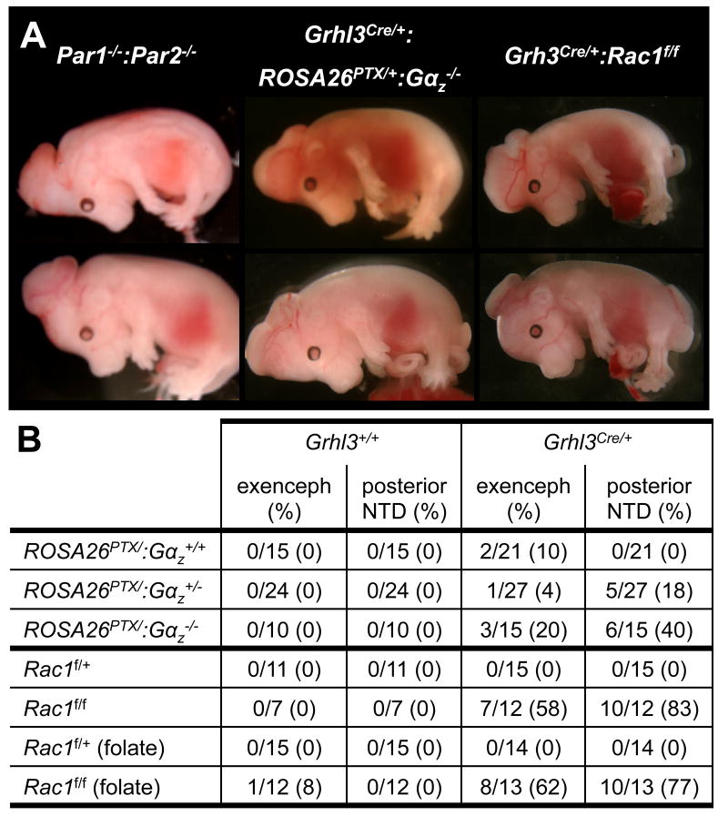
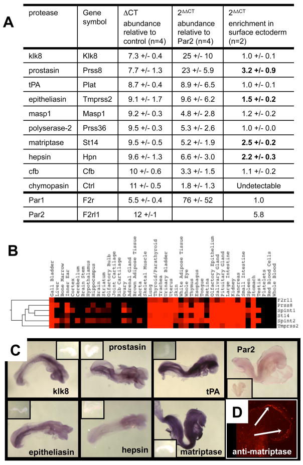
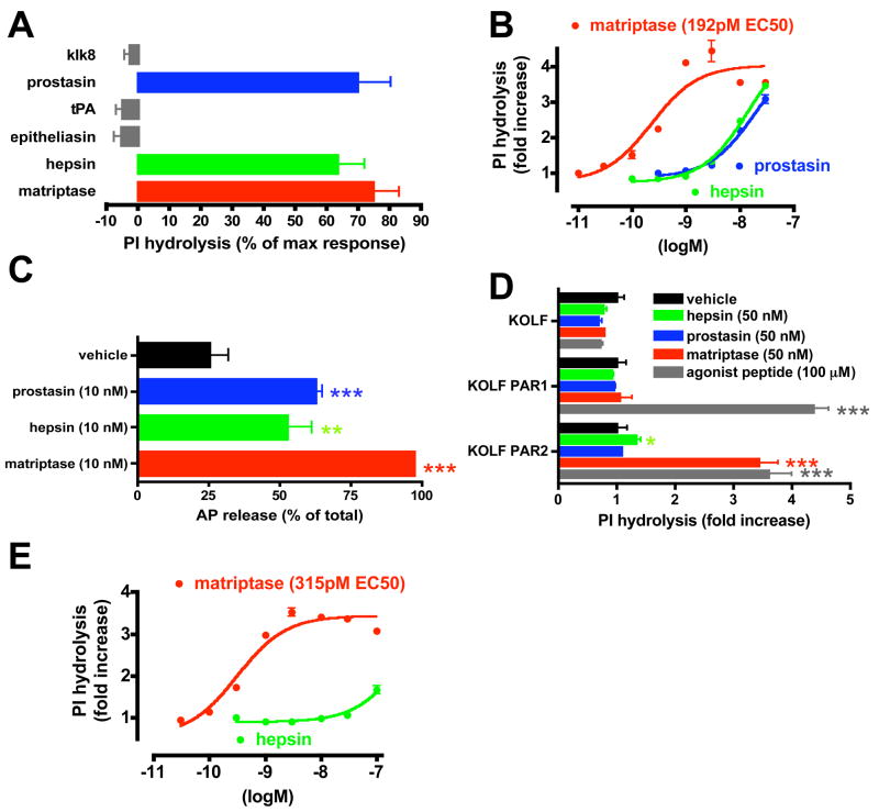
We report an unexpected role for protease signaling in neural tube closure and the formation of the central nervous system. Mouse embryos lacking protease-activated receptors 1 and 2 showed defective hindbrain and posterior neuropore closure and developed exencephaly and spina bifida, important human congenital anomalies. Par1 and Par2 were expressed in surface ectoderm, and Par2 was expressed selectively along the line of closure. Ablation of G(i/z) and Rac1 function in these Par2-expressing cells disrupted neural tube closure, further implicating G protein-coupled receptors and identifying a likely effector pathway. Cluster analysis of protease and Par2 expression patterns revealed a group of membrane-tethered proteases often coexpressed with Par2. Among these, matriptase activated Par2 with picomolar potency, and hepsin and prostasin activated matriptase. Together, our results suggest a role for protease-activated receptor signaling in neural tube closure and identify a local protease network that may trigger Par2 signaling and monitor and regulate epithelial integrity in this context.


Figures

Figure 1
Figure 1. Phenotypes associated with combined deficiency of Par2 and Par1
(A) Three phenotypes exhibited by Par1−/−:Par2/ embryos: mid-gestational cardiovascular failure (top left); late-gestational death associated with edema (top center) and exencephaly (top right). Par1−/−:Par2−/− embryos that survived to birth showed exencephaly and spina bifida with incomplete penetrance (lower left; from left to right: curly tail, spina bifida/curly tail, anencephaly, anencephaly and spina bifida/curly tail). A surviving adult with curly tail is at lower right. (B,C) Viability and presence of exencephaly at various gestational ages (B) and in the presence or absence of an endothelial Par1 transgene (C); # alive (# with exencephaly) is shown. Embryos in (C) were collected at 12.5–14.5 dpc. (D, E) Embryos from Par1+/−:Par2+/− X Par1−/−:Par2−/− crosses were collected at 9.5 dpc, scored for an open HO or closed HC hindbrain neuropore and somite count, then genotyped. To visualize the neuropore, embryos were X-gal stained. (E) Results from embryos in (D) with 15 or more somites. See also Figure S1.
Figure 2
Figure 2. Localized expression of Par2 in surface ectoderm and characterization of a surface ectoderm Cre (Grhl3Cre)
(A–C) Par2 expression at the time of neural tube closure. (A,B) X-gal staining of Par2-lacZ knock-in embryo in whole mount (A) and cross section (B). In situ hybridization of wild-type embryos for Par2 mRNA (C). (D, E) LacZ expression from a Cre-IRES-NLSlacZ cassette inserted into the Grhl3 locus (Grhl3Cre; see Figure S3). (F) In situ hybridization of wild-type embryo for Grhl3 mRNA. Sense controls for C and F are in Figure S2. Embryos were collected at about 9.25 dpc.
Figure 3
Figure 3. Abrogating Gi function and Rac1 expression in surface ectoderm causes neural tube defects
(A) Exencephaly in Par1−/−:Par2−/−, Grhl3Cre/+:ROSA26PTX/+:Gαz−/−, and Grhl3Cre/+:Rac1f/f embryos collected at 14.5 dpc. Spina bifida and curly tail in Grhl3Cre/+:ROSA26PTX/+: Gαz−/− (lower) and Grhl3Cre/+:Rac1f/f (both) embryos. (B) Occurrence of neural tube defects (# with NTD/# live (%)) by genotype at 14.5 dpc. See also Figure S3.
Figure 4
Figure 4. Identification of candidate Par2 activators expressed during neural tube closure
(A) Expression of 125 serine proteases predicted to reside in the extracellular space by real-time PCR of RNA from pooled wild-type embryos collected at 8.5–9.5 dpc. The ten most abundantly expressed relative to housekeeping genes (ΔCT), their abundance relative to Par2 (2ΔΔCT) and their enrichment in surface ectoderm (expression in YFP-positive surface ectoderm cells sorted from dissociated Grhl3Cre+/−:ROSA26YFP/+ (ROSA26 Lox-STOP-Lox YFP) embryos relative to YFP-negative cells from the same embryos (2ΔΔCT)) are shown. (B) Hierarchical cluster analysis was used to compare expression patterns of 125 candidate proteases, 11 select inhibitors and Par2 was measured by real-time PCR of RNA from 44 tissues and circulating cells from adult mice. The cluster containing Par2, matriptase (St14), prostasin (prss8) and their inhibitors Hai-1 (Spint1) and Hai-2 (Spint2) is shown in TreeView; red intensity indicates increased expression. See Document S3 for entire cluster analysis and S1 for experimental procedures. (C) Surface ectoderm expression of mRNA for the indicated candidate proteases and Par2 by whole-mount in situ hybridization of 8.5 dpc embryos; sense controls are in insets. (D) Immunostaining of live 8.5 dpc embryos with E2, an antibody specific for the active form of matriptase. Arrows indicate staining of surface ectoderm overlying the hindbrain neuropore. Close-up is in Figure S4.
Figure 5
Figure 5. Par2 cleavage and activation by membrane-associated proteases
(A,B,E) Protease-triggered phosphoinositide (PI) hydrolysis in response to candidate Par2-activating proteases. (A) PI hydrolysis in HaCaT cultures after addition of soluble recombinant protease domains (50 nM) expressed as % of the response to maximal concentrations of PAR-activating peptides (100 μM TFLLRN + 100 μM SLIGRL). (B) Concentration response to protease domains that showed activity in (A). (C) Alkaline phosphatase (AP) release upon addition of soluble protease domains to HaCaTs transiently expressing an N-terminal AP–Par2 fusion protein. See Figure S5 for cleavage site specificity and PAR selectivity. (D) PI hydrolysis triggered by recombinant soluble protease domains or a maximal concentration (100 μM) of PAR1 or PAR2 peptide agonist (or both peptides for untransfected KOLFs) in KOLFs stably transfected with PAR1 or PAR2. Similar results were obtained in three separate experiments. (E) PI hydrolysis in PAR2-expressing KOLFs, concentration response to matriptase and hepsin.
Figure 6
Figure 6. Evidence for a cascade from prostasin and hepsin to matriptase to Par2 activation
(A) Inhibition of protease-triggered PI hydrolysis in HaCaT by E2 (2 μM), an active site-blocking antibody to matriptase. (B) Par2 cleavage by prostasin in KOLFs transiently transfected with expression vectors for mouse AP-Par2, Hai-1 and full-length matriptase as indicated. AP release in response to addition of soluble recombinant prostasin or matriptase (50nM) was measured. (C) Immunoblot analysis of matriptase processing by prostasin and hepsin in KOLFs transfected with Hai-1 alone or Hai-1 plus matriptase or matriptase active site mutant (asm) expression vectors. Cultures were treated with the soluble recombinant protease domains (50 nM) indicated at top; lysates were immunoblotted with M69 and M24 matriptase antibodies, which recognize the active processed form and total matriptase, respectively. (D) Conversion of endogenously expressed matriptase zymogen to its active form by prostasin and hepsin in HaCaT cultures. Cultures were treated with the indicated soluble recombinant protease domains and cell lysates analyzed as in (C). Tubulin blot served as a loading control. See also Figure S6.

Comment in

References

    1. Auden A, Caddy J, Wilanowski T, Ting SB, Cunningham JM, Jane SM. Spatial and temporal expression of the Grainyhead-like transcription factor family during murine development. Gene Expr Patterns. 2006;6:964–970. - PubMed
    1. Becker RC, Moliterno DJ, Jennings LK, Pieper KS, Pei J, Niederman A, Ziada KM, Berman G, Strony J, Joseph D, et al. Safety and tolerability of SCH 530348 in patients undergoing non-urgent percutaneous coronary intervention: a randomised, double-blind, placebo-controlled phase II study. Lancet. 2009;373:919–928. - PubMed
    1. Benaud C, Dickson RB, Lin CY. Regulation of the activity of matriptase on epithelial cell surfaces by a blood-derived factor. Eur J Biochem. 2001;268:1439–1447. - PubMed
    1. Bhatt AS, Welm A, Farady CJ, Vasquez M, Wilson K, Craik CS. Coordinate expression and functional profiling identify an extracellular proteolytic signaling pathway. Proc Natl Acad Sci U S A. 2007;104:5771–5776. - PMC - PubMed
    1. Briot A, Deraison C, Lacroix M, Bonnart C, Robin A, Besson C, Dubus P, Hovnanian A. Kallikrein 5 induces atopic dermatitis-like lesions through PAR2-mediated thymic stromal lymphopoietin expression in Netherton syndrome. J Exp Med. 2009;206:1135–1147. - PMC - PubMed

Publication types

MeSH terms